. 2019 Apr 5;10(1):1558.
doi: 10.1038/s41467-019-09534-x.

A revised biosynthetic pathway for the cofactor F420 in prokaryotes

Affiliations

A revised biosynthetic pathway for the cofactor F420 in prokaryotes

Ghader Bashiri et al. Nat Commun. .

Abstract

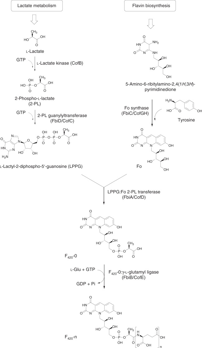
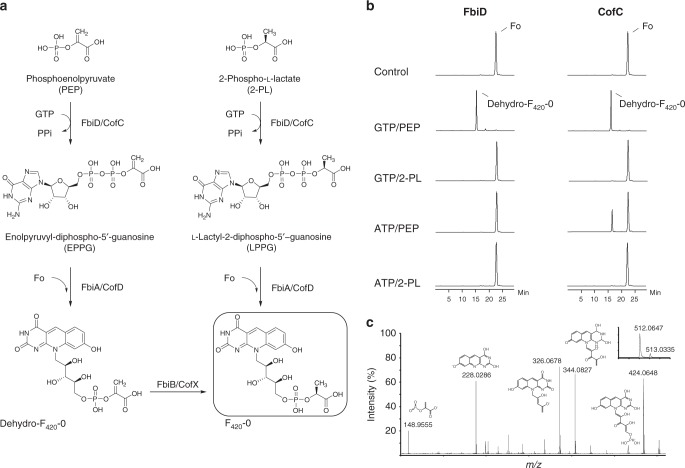
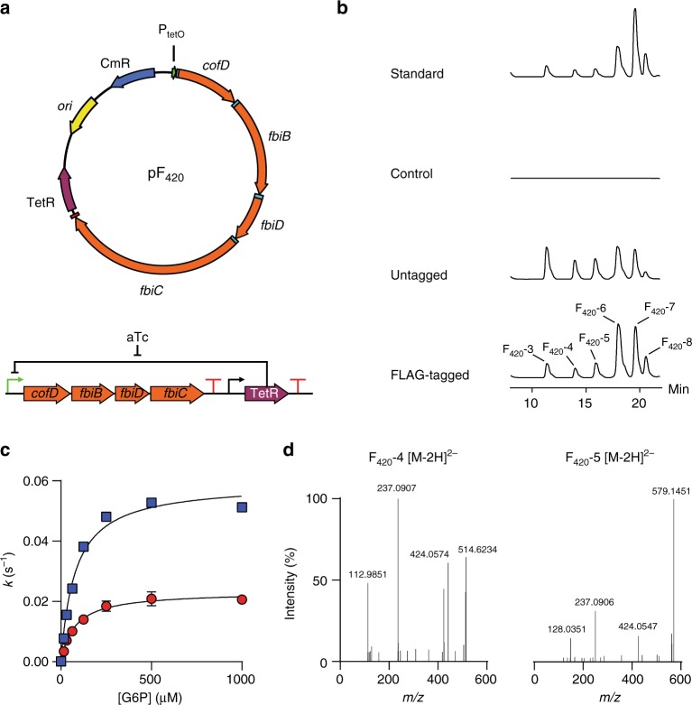
Cofactor F420 plays critical roles in primary and secondary metabolism in a range of bacteria and archaea as a low-potential hydride transfer agent. It mediates a variety of important redox transformations involved in bacterial persistence, antibiotic biosynthesis, pro-drug activation and methanogenesis. However, the biosynthetic pathway for F420 has not been fully elucidated: neither the enzyme that generates the putative intermediate 2-phospho-L-lactate, nor the function of the FMN-binding C-terminal domain of the γ-glutamyl ligase (FbiB) in bacteria are known. Here we present the structure of the guanylyltransferase FbiD and show that, along with its archaeal homolog CofC, it accepts phosphoenolpyruvate, rather than 2-phospho-L-lactate, as the substrate, leading to the formation of the previously uncharacterized intermediate dehydro-F420-0. The C-terminal domain of FbiB then utilizes FMNH2 to reduce dehydro-F420-0, which produces mature F420 species when combined with the γ-glutamyl ligase activity of the N-terminal domain. These new insights have allowed the heterologous production of F420 from a recombinant F420 biosynthetic pathway in Escherichia coli.


Conflict of interest statement

The authors declare no competing interests.

Figures

Fig. 1
Fig. 1
The currently accepted bacterial F420 biosynthesis pathway. The pathway is composed of two branches. In the first branch tyrosine is condensed with the flavin biosynthesis intermediate 5-amino-6-ribitylamino-2,4-(1H,3H)-pyrimidinedione by FbiC/CofGH to produce the riboflavin level chromophore Fo. In the second branch, l-lactic acid is phosphorylated in a guanosine-5ʹ-triphosphate (GTP)-dependent manner by the hypothetical l-lactate kinase CofB to produce 2-phospho-l-lactate (2-PL). This compound is subsequently guanylylated by FbiD/CofC to produce the unstable intermediate l-lactyl-2-diphospho-5ʹ-guanosine (LPPG). The two branches merge as the 2-phospho-l-lactyl moiety of LPPG is transferred to Fo through the action of FbiA/CofD to produce F420-0. Mature F420 is then produced through glutamylation of F420-0 by FbiB/CofE. “Fbi” refers to bacterial proteins, whereas “Cof” represents archaeal ones
Fig. 2
Fig. 2
Phosphoenolpyruvate (PEP) is an intermediate in the formation of dehydro-F420-0. a Production of F420-0 in our revised biosynthesis pathway (left) compared to the currently accepted pathway (right). b Coupled-reaction high-performance liquid chromatography (HPLC) assays showing that both Mtb-FbiD and CofC from Methanocaldococcus jannaschii (Mj-CofC) enzymes use PEP to produce dehydro-F420-0. c Tandem mass spectral identification of dehydro-F420-0. Tandem mass spectrometry (MS/MS) fragmentation of dehydro-F420-0, showing fragment ions with their corresponding structures. The inset displays the observed spectrum of the parent molecule (expected monoisotopic m/z 512.0711 [M − H])
Fig. 3
Fig. 3
Crystal structure of Mtb-FbiD. a Electrostatic surface representation of the Mtb-FbiD structure in complex with phosphoenolpyruvate (PEP), shown as a ball-and-stick model. b The phosphate group of PEP binds to three aspartic acid side chains through two Mg2+ ions (shown in cyan). PEP is shown in 2Fo − Fc omit density contoured at 2.0σ, and drawn as ball-and-stick model. Water molecules are shown as red spheres and hydrogen bond interactions are outlined as dashed lines. c Superposition of Mtb-FbiD (wheat ribbon) and Methanocaldococcus jannaschii (Mj-CofC) (green ribbon), indicating 1.85 Å root mean square difference (rmsd) over 181 superimposed Cα. PEP is shown as a ball-and-stick model
Fig. 4
Fig. 4
Mtb-FbiB catalyzes reduction of dehydro-F420-0. a F420-1 is produced in FbiD:FbiA:FbiB coupled assays in the presence of Fre/FMNH2 and l-glutamate. Tandem mass spectrometry (MS/MS) confirmation of F420-1 in both negative (643.12811, [M − H]) and positive (645.27094, [M + H]+) modes. b Mtb-FbiB is a bifunctional enzyme catalyzing the reduction of dehydro-F420-0 and its poly-glutamylation to form F420-n. c Docking of FMNH2 and dehydro-F420-0 into the crystal structure of FbiB C-terminal domain. The methylene group of the enolpyruvyl moiety sits in a pocket made up of M372 and P289, while the carboxylate hydrogen bonds with R337. The methylene double bond sits planar above the isoalloxazine ring of FMNH2 at an appropriate distance (3.6 Å, shown by dashed line) and oriented for a hydride transfer to the Si face of the methylene bond, accounting for the observed (S)-lactyl moiety of F420
Fig. 5
Fig. 5
Heterologous expression of F420 biosynthesis pathway in E. coli. a Schematic representation of the vector generated for expression of the F420 biosynthesis pathway. b High-performance liquid chromatography-fluorescence detector (HPLC-FLD) traces of E. coli lysates containing F420 biosynthesis constructs as well as a purified standard from M. smegmatis. c Kinetic studies indicate an identical Michaelis constant for glucose-6-phosphate dehydrogenase (FGD) as measured with F420 purified from M. smegmatis (blue squares) and E. coli (red circles). Error bars represent standard deviations. d Fragmentation of F420-4 and F420-5 extracted from E. coli shows a mature F420 production
Fig. 6
Fig. 6
The revised bacterial F420 biosynthesis pathway. The revised pathway is a modified scheme showing that phosphoenolpyruvate (PEP) acts as the substrate for the FbiD/CofC enzymes to produce enolpyruvyl-diphospho-5ʹ-guanosine (EPPG) or enolpyruvyl-diphospho-5ʹ-adenosine (EPPA) (in the case of CofC). The immediate reaction product formed from Fo and EPPG/EPPA is dehydro-F420-0, which is reduced to F420-0 through the newly described reductase activity of the C-terminal domain of FbiB in mycobacteria. A separate enzyme in archaea and some bacteria is expected to catalyze this reduction step (CofX). FbiB/CofE subsequently adds a poly-γ-glutamate tail to form F420. “Fbi” refers to bacterial proteins, whereas “Cof” represents archaeal ones

References

    1. Greening C, et al. Physiology, biochemistry, and applications of F420- and Fo-dependent redox reactions. Microbiol. Mol. Biol. Rev. 2016;80:451–493. doi: 10.1128/MMBR.00070-15. - DOI - PMC - PubMed
    1. Ney B, et al. The methanogenic redox cofactor F420 is widely synthesized by aerobic soil bacteria. ISME J. 2017;11:125–137. doi: 10.1038/ismej.2016.100. - DOI - PMC - PubMed
    1. Walsh C. Naturally occurring 5-deazaflavin coenzymes: biological redox roles. Acc. Chem. Res. 1986;19:216–221. doi: 10.1021/ar00127a004. - DOI
    1. Cheeseman P, Toms-Wood A, Wolfe RS. Isolation and properties of a fluorescent compound, Factor420, from Methanobacterium strain M.o.H. J. Bacteriol. 1972;112:527–531. - PMC - PubMed
    1. Eirich LD, Vogels GD, Wolfe RS. Proposed structure for coenzyme F420 from Methanobacterium. Biochemistry. 1978;17:4583–4593. doi: 10.1021/bi00615a002. - DOI - PubMed

Publication types

MeSH terms

LinkOut - more resources