. 2019 Apr 5;10(1):1577.
doi: 10.1038/s41467-019-09641-9.

Pellino1 regulates reversible ATM activation via NBS1 ubiquitination at DNA double-strand breaks

Affiliations

Pellino1 regulates reversible ATM activation via NBS1 ubiquitination at DNA double-strand breaks

Geun-Hyoung Ha et al. Nat Commun. .

Abstract

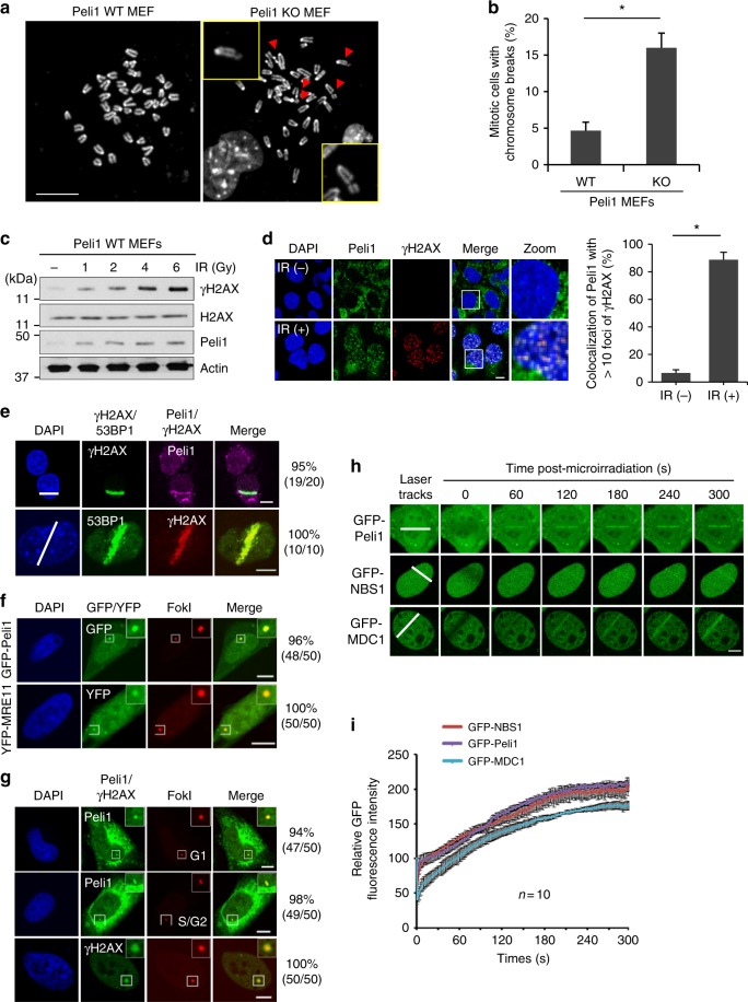
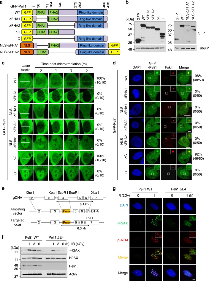
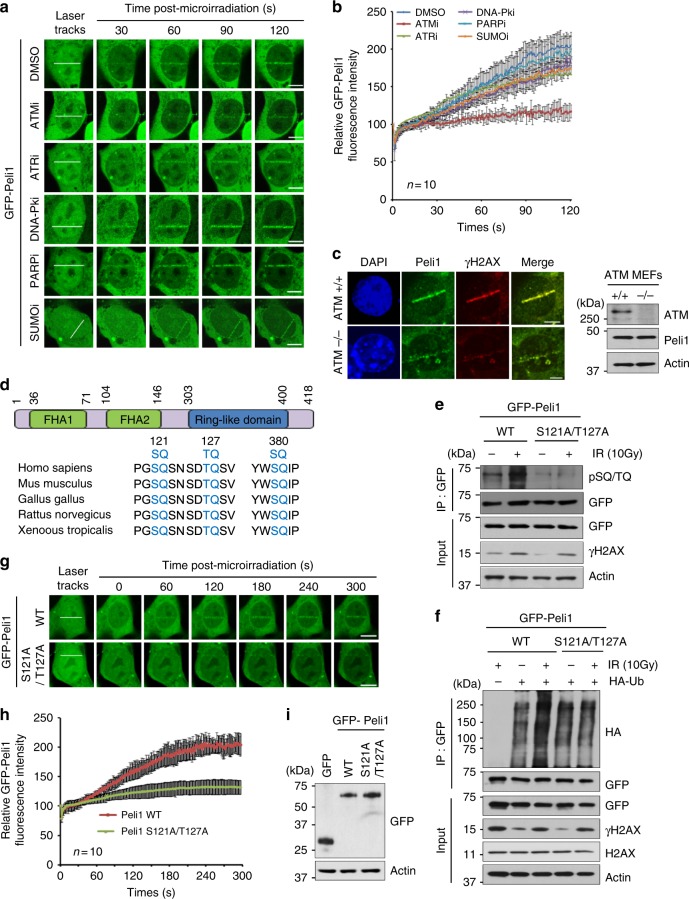
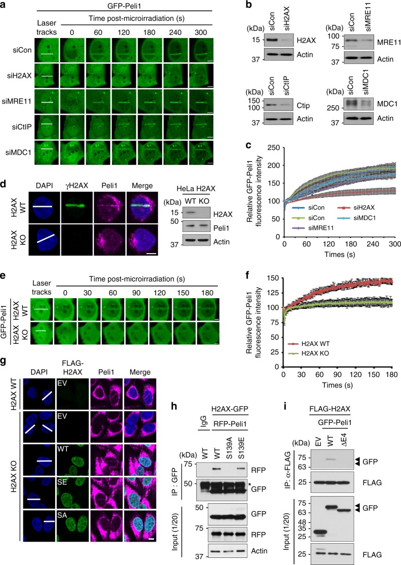
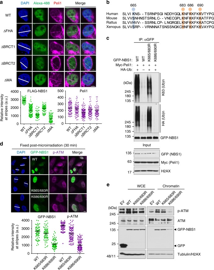
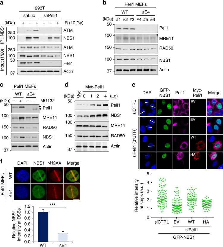
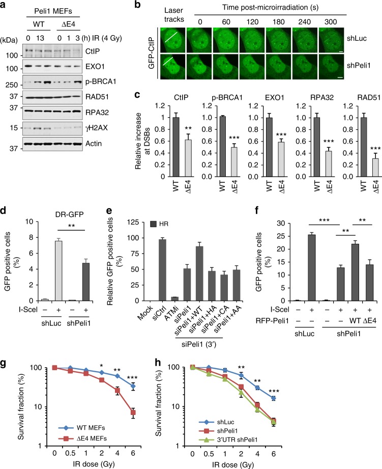
DNA double-strand break (DSB) signaling and repair are critical for genome integrity. They rely on highly coordinated processes including posttranslational modifications of proteins. Here we show that Pellino1 (Peli1) is a DSB-responsive ubiquitin ligase required for the accumulation of DNA damage response proteins and efficient homologous recombination (HR) repair. Peli1 is activated by ATM-mediated phosphorylation. It is recruited to DSB sites in ATM- and γH2AX-dependent manners. Interaction of Peli1 with phosphorylated histone H2AX enables it to bind to and mediate the formation of K63-linked ubiquitination of NBS1, which subsequently results in feedback activation of ATM and promotes HR repair. Collectively, these results provide a DSB-responsive factor underlying the connection between ATM kinase and DSB-induced ubiquitination.


Conflict of interest statement

The authors declare no competing interests.

Figures

Fig. 1
Fig. 1
Peli1 is recruited to DNA double-strand break sites. a Mitotic spreads of primary Peli1 wild-type (WT) and Peli1 knockout (KO). MEF cells were arrested in metaphase with 0.1 mg/ml of colchicine, fixed, and visualized by DAPI staining. Red arrow designates chromatid and chromosome breaks. Scale bars, 5 μm. b Quantification of the number of chromatid and chromosome breaks per cell evaluated in 50 cell metaphase spreads. Student’s t-test was used for statistical analyses. c Peli1 WT MEF cells were treated with IR at different doses (0, 1, 2, 4, and 6 Gy). At 1 hr after IR, cells were collected and then subjected to immunoblotting with Peli1, γH2AX, H2AX, and actin antibodies. d Peli1 WT MEFs were either control treated or treated with IR (10 Gy) and allowed to recover for 1 h before processing for Peli1 and γH2AX immunostaining (left). Scale bars, 10 μm. Quantitative analysis for colocalization of Peli1 with >10 foci of γH2AX without IR or with IR. Plotted values represent mean ± SEM of more than 300 individual cells. Student’s t-test was used for statistical analyses. e Peli1 WT MEF cells were subjected to laser micro-irradiation. After 10 min, cells were co-stained with γH2AX or 53BP1 and Peli1 antibodies. The white lines with DAPI staining indicated laser stripes. Scale bars, 10 μm. f mCherry-LacI-FokI expressing plasmid was cotransfected with GFP-Peli1 (upper panels) or YFP-MRE11 (positive control, lower panels) into U2OS-DSB reporter cells. Scale bars, 10 μm. g mCherry-LacI-FokI plasmid was transfected into U2OS-DSB reporter cells. At 48 h post transfection, cells were immunostained with Peli1 or γH2AX antibodies. The number of FokI focus determines the cell cycle phase. Single FokI (upper panel) represents G1 phase while double FokI represents S/G2 phase of cell cycle. Scale bars, 10 μm. h, i U2OS cells were transfected with GFP-Peli1, GFP-NBS1, or GFP-MDC1 expressing plasmid. At 48 h, cells were subjected to laser micro-irradiation. Laser stripes were examined at indicated time point (h). The intensity of each laser stripe was determined by averaging values from ten cells at each time point and graphed (i). Scale bars, 10 μm
Fig. 2
Fig. 2
Deletion of Peli1 FHA domains impaired DNA damage response. a Diagrams of Peli1 WT, deletion, and NLS-fused deletion mutants. b 293T cells were transfected with plasmids encoding GFP-Peli1 WT, each of deletion and NLS-fused mutants. At 48 h post transfection, cells were collected and cell lysates were analyzed by immunoblotting with anti-GFP and anti-tubulin antibodies. c U2OS cells were transfected with GFP-Peli1 WT or each mutant and treated with BrdU (10 μM) for 30 h followed by laser micro-irradiation. Scale bar, 10 μM. d mCherry-LacI-FokI and GFP-fused Peli1 WT or mutants were cotransfected into U2OS-DSB reporter cells (U2OS 2–6–3). At 48 h, cells were fixed and visualized by confocal microscopy. Scale bars, 10 μm. e Targeting strategy to generate PELI1 mutant mice in which coding for exon 4 was replaced with a puromycin cassette gene, resulting in Peli1 E4 truncated mice. f Peli1 WT and E4 truncated MEFs were treated with IR (4 Gy) as indicated. Cells were collected and then subjected to immunoblotting with Peli1, γH2AX, H2AX, and actin antibodies. g Peli1 WT and E4 truncated MEFs were treated with IR (2 Gy) and allowed to recover for 1 h before processing for γH2AX and phospho-ATM (p-Ser1981) immunostaining. Scale bars, 10 μm
Fig. 3
Fig. 3
Peli1 is phosphorylated by ATM that translocate to DNA damage sites. a, b U2OS cells transiently expressing GFP-Peli1 were pre‐treated with ATM (KU55933, ATMi, 10 μM), ATR (VE-831, ATRi, 10 μM), DNA‐PK (KU57788, DNA‐PKi, 10 μM), PARP (PJ34, PARPi, 100 μM), and SUMO (2Dd-08, SUMOi, 50 μM) inhibitors for 1 h. Cells were subjected to laser micro-irradiation. Laser stripes were examined at the indicated time point (a). Scale bars, 10 μm. The intensity of each laser stripe was determined by averaging values from ten cells at each time point and graphed (b). c ATM WT and KO MEF cells were subjected to laser micro-irradiation. Co-immunostaining with γH2AX and Peli1 antibodies at laser-induced DNA lesions (10 min after laser micro-irradiation). Scale bar, 10 μM. d Schematic showing potential Peli1 phosphorylation sites by ATM/ATR kinases. e 293T cells were cotransfected with GFP-Peli1 WT and S121A/T127A mutant. At 48 h post transfection, cells were treated with IR (10 Gy). At 30 min after IR, cells were collected and immunoprecipitated with an anti-GFP antibody. GFP-Peli1 protein complexes were subjected to immunoblotting with anti-pSQ/TQ, anti-GFP, anti-γH2AX, and anti-actin antibodies. f 293T cells were cotransfected with GFP-Peli1 WT and S121A/T127A mutant with or without HA-Ub. At 36 h post transfection, cells were treated with IR (10 Gy) and 30 min later cells were collected and immunoprecipitated with an anti-GFP antibody. GFP-Peli1 protein complexes were subjected to immunoblotting with indicated antibodies. g, h U2OS cells were transfected with plasmids encoding GFP-Peli1 WT or S121A/T127A mutant. At 48 h, cells were subjected to laser micro-irradiation and laser stripes were examined at indicated time point. Scale bar, 10 μM (g). The intensity of each laser stripe was determined by averaging values from ten cells at each time point and graphed (h). i U2OS cells were transfected with plasmids encoding GFP-Peli1 WT or S121A/T127A mutant. At 48 h post transfection, cells were collected and analyzed by immunoblotting with anti-GFP and anti-actin antibodies
Fig. 4
Fig. 4
γH2AX-mediated accumulation of Peli1 at DNA damage sites. a, b Mobilization kinetics of GFP-Peli1 to sites of DNA damage. U2OS cells were cotransfected with plasmids encoding GFP-Peli1 and indicated siRNAs. After 48 h, cells were subjected to laser micro-irradiation and laser stripes were examined at indicated time point. Scale bar, 10 μM. U2OS cells were cotransfected with plasmids encoding GFP-Peli1 and indicated siRNAs (a). After 48 h, cell lysates were analyzed by immunoblotting with indicated antibodies (b). c The intensity of each laser stripe was determined by averaging values from ten cells at each time point and graphed. d H2AX WT and KO HeLa cells were subjected to laser micro-irradiation. Colocalization of Peli1 and γH2AX at laser-induced DNA lesions (10 min after laser micro-irradiation; left panels) and western blotting analysis (right panels) are shown. Scale bar, 10 μM. e, f H2AX WT and KO HeLa cells were transfected with GFP-Peli1. At 48 h, cells were subjected to laser micro-irradiation. Laser stripes were examined at the indicated time point (e). The intensity of each laser stripe was determined by averaging values from ten cells at each time point and graphed (f). g H2AX WT and KO HeLa cells were transfected with FLAG empty vector (EV), FLAG-H2AX WT, S139A, or S139E mutant. At 48 h post transfection, cells were subjected to laser micro-irradiation. After 10 min post micro-irradiation, cells were fixed and immunostained with indicated antibodies. Scale bar, 10 μM. h H2AX HeLa cells were cotransfected with H2AX-GFP WT, S139A, or S139E mutant. After 48 h, cells were collected and immunoprecipitated with an anti-GFP antibody. These protein complexes were subjected to immunoblotting with indicated antibodies. i 293T cells were cotransfected with FLAG-H2AX WT and GFP empty vector (EV), GFP-Peli1 WT, or GFP-Peli1 E4 truncated. At 48 h post transfection, cells were collected and immunoprecipitated with an anti-FLAG antibody. These protein complexes were subjected to immunoblotting with antibodies shown
Fig. 5
Fig. 5
Peli1-mediated ATM activation and NBS1 interaction upon DNA damage. a Peli1 WT and ΔE4 MEF cells were treated with 4 Gy IR. At indicated time, cells were collected and then subjected to immunoblotting. b Peli1 WT and E4 truncated MEF cells were micro-irradiated, fixed after 10 min, and immunostained with anti-p-ATM antibody (left panels). Mean levels of p-ATM accumulation at sites of laser tracks were quantified using Image J software and plotted as indicated (right panels). Data show mean ± SEM; n = 20 cells. Student’s t-test was used for statistical analyses. Scale bars, 10 μm. c Peli1 E4 truncated MEFs were transfected with a control pBabe or pBabe-Peli1 (Myc-Peli1) retrovirus and treated with IR (4 Gy). At 1 h post-IR, cells were collected and immunoblotted with anti-Peli1, anti-p-ATM, anti-ATM, and anti-actin antibodies. d Peli1 E4-truncated MEFs were transfected with pBabe or pBabe-Peli1 (Myc-Peli1) retrovirus for 24 h and then treated with IR (4 Gy). At 1 h post-IR, cells were fixed and immunostained with anti-p-ATM antibodies. Scale bars, 10 μm. e 293T cells were transfected with TAP (control) or TAP-Peli1 (Flag-tagged Peli1). At 36 h, cells were treated with or without IR (4 Gy). At 30 min post-IR, cells were collected and isolated through S-tag pull-down assay. Bound proteins were immunoblotted with indicated antibodies. f IR-treated or non-treated 293T cell lysates were immunoprecipitated with anti-NBS1 or anti-Peli1 antibody and immunoblotted with antibodies indicated. γH2AX was used as a positive marker of IR in input panel
Fig. 6
Fig. 6
Phosphorylation-mediated Peli1 activates NBS1 ubiquitination in vivo. a 293T cells were transfected with GFP-Peli1 WT, GFP-Peli1 C-terminal deletion, or C-terminal mutant in combination with HA-Ub and Myc-NBS1. At 36 h post transfection, cells were collected and immunoprecipitated with an anti-Myc antibody. NBS1 protein complexes were subjected to immunoblotting with anti-HA and anti-Myc antibodies. b Diagram of consensus motifs of SQ/TQ and RING domain of Peli1 protein in eukaryotes. c 293T cells were transfected with GFP-Peli1 WT, H313A (HA), C336A (CA), or S121A/T127A (AA) together with HA-Ub. At 48 h, cells were treated with IR. Cell lysates were immunoprecipitated with anti-GFP antibody. Autoubiquitination of GFP-Peli1 constructs was detected with anti-HA antibody. d, e K63-linked ubiquitination of NBS1 by Peli1. siRNA-targeting Peli1 (d) and plasmid encoding GFP-Peli1 (e) were transfected into 293T cells, respectively. Cell lysates were immunoprecipitated with anti-NBS1 antibody. NBS1 ubiquitination was detected by anti-NBS1-, anti-HA-, and K63-specific antibodies. f 293T cells were transfected with GFP-Peli1, FLAG-NBS1, and HA-Ub. At 24 h post transfection, siRNAs targeting UBC13 #1 and #2 (U13), or control (CL) was reintroduced into cells. After 48 h, cells were immunoprecipitated with anti-FLAG antibody. NBS1 ubiquitination was detected with anti-FLAG antibody
Fig. 7
Fig. 7
Peli1 ubiquitinates NBS1 at lysine 686 and 690 that regulate NBS1 and active ATM retention at damaged sites. a FLAG-NBS1 WT and truncated mutants were transfected into U2OS cells. At 48 h post transfections, cells were microirradated and fixed at 30 min post micro-irradiation. Recruitments of Peli1 and FLAG-NBS1 constructs were evaluated with anti-FLAG and anti-Peli1 antibodies (upper panels). Quantitative analysis of FLAG-NBS1 constructs and Peli1 at laser stripes was also performed. The intensity of each constructs at laser stripes was determined by averaging values from 20 cells and graphed (lower panels). Scale bars, 10 μm. b Comparison of conserved lysine residues of C-terminal NBS1 in eukaryotes. c 293T cells were transfected with GFP-NBS1 WT and lysine-dead (K665/683R and K686/690R) mutants in combination with Myc-Peli1 WT and HA-Ub. Immunoprecipitated NBS1 with anti-GFP antibody was detected with anti-HA and anti-K63 antibodies. d Cells transfected with GFP-Peli1 WT, K665/683R, and K686/690R mutants were subjected to micro-irradiation at 24 h post transfection with endogenous NBS1 3′-UTR-targeting siRNA. At 30 min post micro-irradiation, micro-irradiated cells were fixed and immunostained with anti-p-ATM antibody (upper panels). Quantitative analysis of GFP-NBS1 constructs and p-ATM at laser stripes was also performed. The intensity at laser stripes was determined by averaging values from 20 cells and graphed (lower panels). Scale bars, 10 μm. e 293T cells were transfected with GFP-NBS1 WT, K665/683R, or K686/690R mutants. At 48 h post transfection, DSB-mimetic drug phleomycin (50 μg/ml) was used to treat cells. After washing out, whole cell extracts and chromatin fraction were collected 30 min later. Indicated antibodies were used to detect signals
Fig. 8
Fig. 8
Peli1-mediated NBS1 ubiquitination stabilizes the Mre11, RAD50, and NBS1 (MRN) complex. a 293T cells were transfected with shLuc or Peli1 and treated with or without IR (10 Gy). At 30 min post-IR, cells were collected and immunoprecipitated with an anti-NBS1 antibody. NBS1 protein complexes were subjected to immunoblotting. b Immunoblotting of MRN complex components in Peli1 WT and E4 truncated MEF cells using antibodies indicated. c Peli1 WT and E4 truncated MEF cells were treated with proteasome inhibitor MG132 (10 μM) for 6 h. Cells were subsequently subjected to immunoblotting. d 293T cells were transfected with Myc-Peli1 expression plasmid. At 48 h post transfection, cells were collected and cell lysates were analyzed by immunoblotting with antibodies indicated. e siRNA-targeting control (CTRL) or endogenous Peli1 (3′-UTR) was transfected into U2OS cells in combination with GFP-NBS1. U2OS cells reintroduced with empty vector (EV), Myc-Peli1 WT, or RING mutant (HA) were subjected to micro-irradiation at 48 h. At 30 min post micro-irradiation, cells were fixed and immunostained with anti-Peli1 and anti-Myc antibodies. The intensity of GFP-NBS1 at laser stripes was determined by averaging values from 10 to 20 cells and graphed. Scale bars, 10 μm. f Peli1 WT and E4 truncated MEF cells were micro-irradiated with a UV laser, fixed after 10 min, and immunostained with anti-NBS1 and anti-γH2AX antibodies (upper panel). Scale bar, 10 μM. Mean levels of NBS1 accumulation at sites of laser tracks were quantified (lower panel). Student’s t-test was used for statistical analyses
Fig. 9
Fig. 9
Peli1 involves DNA-end resection for HR repair. a Peli1 WT and E4 truncated MEF cells were treated with IR (4 Gy). At 0, 1, and 3 h post-IR, cells were collected and immunoblotted. b U2OS cells stably expressing GFP-CtIP were transfected with shLuc or shPeli1. After 48 h, cells were subjected to laser micro-irradiation. Laser stripes were examined at indicated time points. Scale bars, 10 μm. c Peli1 WT and E4 truncated MEF cells were micro-irradiated at 10 min for CtIP and p-BRCA1, at 30 min for Exo1, RPA32, and Rad51. Fixed cells were immunostained with indicated antibodies. All data are presented as mean values ± SEM of three independent experiments; n = 20 cells. Student’s t-test was used for statistical analyses. d, e U2OS HR reporter cells were transfected with shRNA targeting Peli1 or control (d), or siRNA-targeting Peli1 3′-UTR region and Myc-Peli1 WT, RING mutants (HA and CA), and phospho-dead mutant (AA) in combination as indicated (e). After 24 h, I-SceI nuclease-expressing vector was transfected into reporter cells. After 48 h, GFP-positive cells were calculated by FACS. Student’s t-test was used for statistical analyses. An ATM inhibitor (ATMi) was used as a HR positive control (e). f Twenty-four hours after transfection of shLuc or shPeli1 vector in 293T cells expressing pDR-GFP, I-SceI expression vector was further transfected into these cells to generate a DSB within Sce-GFP. FACS analysis was then performed to quantify HR-repaired GFP-positive cells. Student’s t-test was used for statistical analyses. g Peli1 WT or E4 truncated MEF cells were treated with indicated doses of IR. Clonogenic survival assays were performed three independent experiments and survival curves were generated. Results are presented as mean ± SEM. Student’s t-test was used for statistical analyses. h U2OS cells silenced with control (shLuc) or Peli1 (shPeli1 or 3′-UTR shPeli1) were treated with various doses of IR and survival rate of these cells was determined by colony formation assay. Results are mean ± SEM of three independent experiments. Student’s t-test was used for statistical analyses
Fig. 10
Fig. 10
Model of Peli1-mediated DNA damage signaling and HR repair. Peli1 recruitment to DSB sites is triggered by ATM-mediated phosphorylation and directly binds to phosphorylated histone H2AX (Step I). Once recruited, Peli1 interacts with NBS1 and catalyzes K63-linked ubiquitination of NBS1, leading to further activation of ATM, which reinforces DNA-end resection and HR repair (Step II)

References

    1. Schwertman P, Bekker-Jensen S, Mailand N. Regulation of DNA double-strand break repair by ubiquitin and ubiquitin-like modifiers. Nat. Rev. Mol. Cell Biol. 2016;17:379–394. doi: 10.1038/nrm.2016.58. - DOI - PubMed
    1. Polo SE, et al. Regulation of DNA-end resection by hnRNPU-like proteins promotes DNA double-strand break signaling and repair. Mol. Cell. 2012;45:505–516. doi: 10.1016/j.molcel.2011.12.035. - DOI - PMC - PubMed
    1. Durocher D, Jackson SP. DNA-PK, ATM and ATR as sensors of DNA damage: variations on a theme? Curr. Opin. Cell Biol. 2001;13:225–231. doi: 10.1016/S0955-0674(00)00201-5. - DOI - PubMed
    1. Abraham RT. Cell cycle checkpoint signaling through the ATM and ATR kinases. Genes Dev. 2001;15:2177–2196. doi: 10.1101/gad.914401. - DOI - PubMed
    1. Stracker TH, Usui T, Petrini JH. Taking the time to make important decisions: the checkpoint effector kinases Chk1 and Chk2 and the DNA damage response. DNA Repair (Amst.) 2009;8:1047–1054. doi: 10.1016/j.dnarep.2009.04.012. - DOI - PMC - PubMed

Publication types

MeSH terms