Review
. 2019 May;15(5):273-287.
doi: 10.1038/s41584-019-0202-1.

Spontaneous dog osteoarthritis - a One Medicine vision

Affiliations
Review

Spontaneous dog osteoarthritis - a One Medicine vision

Richard L Meeson et al. Nat Rev Rheumatol. 2019 May.

Abstract

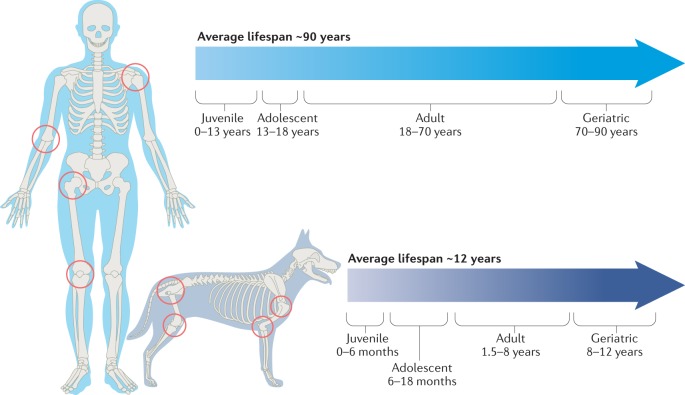
Osteoarthritis (OA) is a global disease that, despite extensive research, has limited treatment options. Pet dogs share both an environment and lifestyle attributes with their owners, and a growing awareness is developing in the public and among researchers that One Medicine, the mutual co-study of animals and humans, could be beneficial for both humans and dogs. To that end, this Review highlights research opportunities afforded by studying dogs with spontaneous OA, with a view to sharing this active area of veterinary research with new audiences. Similarities and differences between dog and human OA are examined, and the proposition is made that suitably aligned studies of spontaneous OA in dogs and humans, in particular hip and knee OA, could highlight new avenues of discovery. Developing cross-species collaborations will provide a wealth of research material and knowledge that is relevant to human OA and that cannot currently be obtained from rodent models or experimentally induced dog models of OA. Ultimately, this Review aims to raise awareness of spontaneous dog OA and to stimulate discussion regarding its exploration under the One Medicine initiative to improve the health and well-being of both species.


Conflict of interest statement

The authors declare no competing interests.

Figures

Fig. 1
Fig. 1. Osteoarthritis in dogs and humans.
The most common locations for osteoarthritis in dogs include the knee, hip, shoulder and elbow, which are shown with their homologous equivalent in humans. The average lifespan of a large-breed dog is ~12 years, with a proportionately longer time spent in old age than in a typical human lifespan.
Fig. 2
Fig. 2. Analogous disease in dogs and humans.
Similarities in the aetiopathology of dog (yellow) and human (blue) forms of osteoarthritis (OA) are shown for developmental vascular OA of the hip (Legg-Calvé-Perthes disease (LCPD) in dogs and LCPD and avascular necrosis of the femoral head in humans), developmental joint instability OA (canine hip dysplasia (CHD) in dogs and developmental dysplasia of the hip (DDH) in humans) and acquired adult joint instability OA (knee anterior cruciate ligament (ACL) rupture in humans and cranial cruciate ligament (CCL) rupture in dogs). IGF1, insulin-like growth factor 1; SNP, single nucleotide polymorphism.
Fig. 3
Fig. 3. Hip dysplasia and hip osteoarthritis in dogs and humans.
Hip dysplasia commonly occurs in juvenile large-breed dogs, such as Golden Retrievers, and can result in osteoarthritis (OA) in later years. a | Radiograph of an adult dog with severe hip dysplasia and luxated hips. The image was acquired as a ventrodorsal extended limb radiograph. b | Radiograph of a human infant with a dysplastic luxated left hip. c | Radiograph of a 3-month-old dog with bilateral dysplastic and subluxated hips. The image was acquired in a dorsolateral subluxation screening position. d,e | Radiographs of the hip joints from a middle-aged male human with primary OA (part d) and a middle-aged female large-breed dog with secondary OA subsequent to hip dysplasia (part e). Both human and dog osteoarthritic hips show evidence of advanced new bone formation and sclerosis of the acetabulae and the femoral head and neck region. Part b courtesy of R. T. Loder, Riley Children’s Hospital, USA.
Fig. 4
Fig. 4. Cruciate ligament rupture and knee osteoarthritis in dogs and humans.
Cranial cruciate ligament (CCL; analogous to the human anterior cruciate ligament (ACL)) rupture occurs in most breeds of dog but is particularly common in young large-breed dogs, such as Labrador Retrievers, and older small-breed dogs. a | Post-mortem dog knee with a healthy CCL (arrow). b | Post-mortem dog knee with a spontaneously degenerated, partially ruptured CCL (arrow shows degeneration of the craniomedial (anteromedial) band). c | Mediolateral radiograph of a healthy dog knee that shows no signs of effusion, sclerosis, soft tissue thickening or osteophytosis. d | Mediolateral radiograph of a dog knee with a ruptured CCL and osteoarthritic changes. Radiographic joint effusion and new bone formation associated with the distal pole of the patella, the tibial plateau, trochlear ridges and the insertion of the CCL are evident. e | Proton density turbo spin echo sequence sagittal MRI of a healthy human knee. f | T1-weighted sagittal MRI of a healthy dog knee showing the CCL in part and the caudal cruciate ligament in full. g | MRI of an adolescent human knee with a ruptured ACL (arrows indicate the space without an intact ligament). h | MRI of a dog stifle joint with a ruptured CCL (arrow indicates space without a CCL). Part e courtesy of K. Chappell, Imperial College London, UK.
Fig. 5
Fig. 5. Shoulder osteochondritis dissecans in dogs.
Shoulder osteochondritis dissecans lesions predominantly occur in adolescent large-breed dogs. a,b | Lateral radiograph (part a) showing a classical mineralized flap over the caudal region of the humeral head, which can also be seen in a transverse CT (part b) of the same shoulder with associated subchondral sclerosis (arrows mark the lesion). c | Osteochondritis dissecans flap being removed arthroscopically.
Fig. 6
Fig. 6. Avascular necrosis of the femoral head (Legg-Calvé-Perthes disease) in dogs.
Legg-Calvé-Perthes disease (LCPD) occurs predominantly in the hip joints of small-breed dogs. a | Excised femoral head with an abnormal articular surface morphology and a central dark line indicating an articular surface defect. b | Ventrodorsal radiograph of a dog hip joint showing typical LCPD focal lucencies and new bone formation, as seen in advanced lesions.

References

    1. Murphy L, Helmick CG. The impact of osteoarthritis in the United States: a population-health perspective: a population-based review of the fourth most common cause of hospitalization in U.S. adults. Orthop. Nurs. 2012;31:85–91. doi: 10.1097/NOR.0b013e31824fcd42. - DOI - PubMed
    1. March, L. et al. Osteoarthritis: a serious disease. OARSI.orghttps://www.oarsi.org/sites/default/files/docs/2016/oarsi_white_paper_oa... (2016).
    1. Little CB, Zaki S. What constitutes an ‘animal model of osteoarthritis’ – the need for consensus? Osteoarthr. Cartil. 2012;20:261–267. doi: 10.1016/j.joca.2012.01.017. - DOI - PubMed
    1. Pond MJ, Nuki G. Experimentally-induced osteoarthritis in the dog. Ann. Rheum. Dis. 1973;32:387–388. doi: 10.1136/ard.32.4.387. - DOI - PMC - PubMed
    1. Stockwell RA, Billingham ME, Muir H. Ultrastructural changes in articular cartilage after experimental section of the anterior cruciate ligament of the dog knee. J. Anat. 1983;136:425–439. - PMC - PubMed