. 2019 Apr 5;18(1):82.
doi: 10.1186/s12943-019-1016-0.

Long non-coding RNA GBCDRlnc1 induces chemoresistance of gallbladder cancer cells by activating autophagy

Affiliations

Long non-coding RNA GBCDRlnc1 induces chemoresistance of gallbladder cancer cells by activating autophagy

Qiang Cai et al. Mol Cancer. .

Erratum in

Abstract

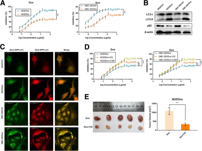
Background: Gallbladder cancer is the most common biliary tract malignancy and not sensitive to chemotherapy. Autophagy is an important factor prolonging the survival of cancer cells under chemotherapeutic stress. We aimed to investigate the role of long non-coding RNAs (lncRNAs) in autophagy and chemoresistance of gallbladder cancer cells.

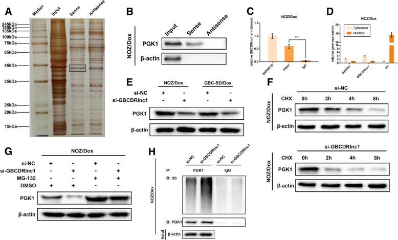
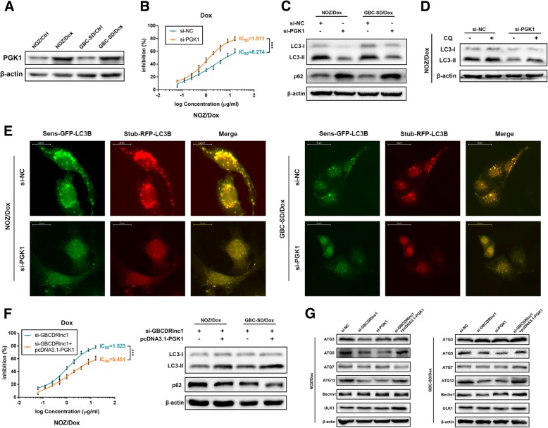
Methods: We established doxorubicin (Dox)-resistant gallbladder cancer cells and used microarray analysis to compare the expression profiles of lncRNAs in Dox-resistant gallbladder cancer cells and their parental cells. Knockdown or exogenous expression of lncRNA combined with in vitro and in vivo assays were performed to prove the functional significance of lncRNA. The effects of lncRNA on autophagy were assessed by stubRFP-sensGFP-LC3 and western blot. We used RNA pull-down and mass spectrometry analysis to identify the target proteins of lncRNA.

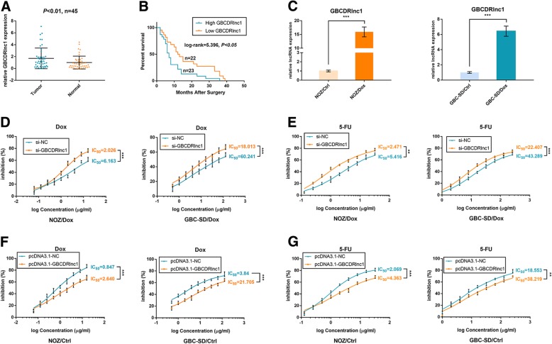
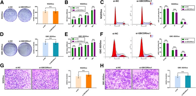
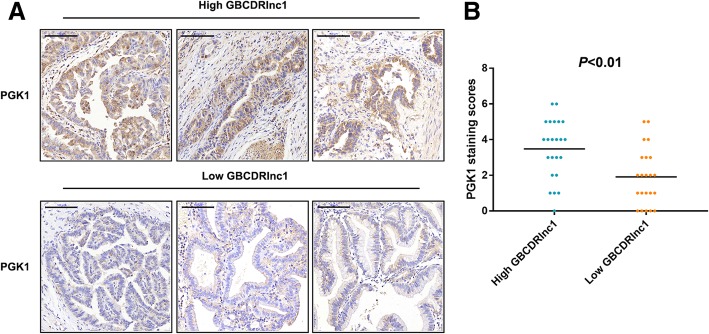
Results: The drug-resistant property of gallbladder cancer cells is related to their enhanced autophagic activity. And we found a lncRNA ENST00000425894 termed gallbladder cancer drug resistance-associated lncRNA1 (GBCDRlnc1) that serves as a critical regulator in gallbladder cancer chemoresistance. Furthermore, we discovered that GBCDRlnc1 is upregulated in gallbladder cancer tissues. Knockdown of GBCDRlnc1, via inhibiting autophagy at initial stage, enhanced the sensitivity of Dox-resistant gallbladder cancer cells to Dox in vitro and in vivo. Mechanically, we identified that GBCDRlnc1 interacts with phosphoglycerate kinase 1 and inhibits its ubiquitination in Dox-resistant gallbladder cancer cells, which leads to the down-regulation of autophagy initiator ATG5-ATG12 conjugate.

Conclusions: Our findings established that the chemoresistant driver GBCDRlnc1 might be a candidate therapeutic target for the treatment of advanced gallbladder cancer.

Keywords: Autophagy; Chemoresistance; Gallbladder cancer; PGK1; lncRNA GBCDRlnc1.


Conflict of interest statement

Ethics approval and consent to participate

Written informed consent was obtained from all patients in accordance with the Declaration of Helsinki. This study was approved by the Human Ethics Committee of Xinhua Hospital (Shanghai JiaoTong University School of Medicine, Shanghai, China). All experiments on the participants in this study were performed in accordance with the relevant guidelines and regulations. All animal experiments were approved by the Animal Care and Use committee of Xinhua Hospital.

Consent for publication

All authors agree the publication of this study.

Competing interests

The authors declare that they have no competing interests.

Publisher’s Note

Springer Nature remains neutral with regard to jurisdictional claims in published maps and institutional affiliations.

Figures

Fig. 1
Fig. 1
The drug-resistant property of Dox-resistant gallbladder cancer cells is associated with their enhanced autophagic activity. a The sensitivities of Dox-resistant gallbladder cancer cells and their parental cells with Dox were determined by CCK-8 assay. b The protein levels of LC3 and p62 in Dox-resistant gallbladder cancer cells and their parental cells were determined by western blot assay. c Dox-resistant gallbladder cancer cells and their parental cells that stably express the stubRFP-sensGFP-LC3 fusion protein were established and observed by the fluorescence microscope. d The sensitivities of Dox-resistant gallbladder cancer cells and their parental cells under different treatments with Dox were determined by CCK-8 assay. e The nude mice carrying tumors from NOZ/Dox under Dox with or without CQ were shown. Average tumor volume for each group was calculated. The mean ± SD of triplicate experiments were plotted, ***P < 0.001
Fig. 2
Fig. 2
LncRNA expression profile in NOZ/Dox cells. a The scatter plot was used for assessing the variation in lncRNA expression between NOZ/Dox and NOZ/Ctrl cells. The values of x and y axes in the scatter plot were the normalized signal values of the samples (log2 scaled). The gray dotted lines are fold-change lines. The lncRNAs above the top gray dotted line and below the bottom gray dotted line indicated more than 2.0-fold change of lncRNAs between the two compared samples. The volcano plot was constructed using fold-change values and P-values. The vertical lines correspond to 2.0-fold up and down, respectively, and the horizontal line represents a P-value of 0.05. The green and red point in the plot represents the differentially expressed lncRNAs with statistical significance. b The cluster heat map showed the differentially expressed lncRNAs over 2.0-fold change. Red colour indicates high expression level, and green colour indicates low expression level. c The top 10 upregulated lncRNAs and top 10 downregulated lncRNAs. The arrow indicates GBCDRlnc1
Fig. 3
Fig. 3
GBCDRlnc1 is upregulated in gallbladder cancer tissues and promotes chemoresistance of gallbladder cancer cells in vitro. a Relative expression of GBCDRlnc1 in gallbladder cancer tissues and neighboring noncancerous tissues was detected by qRT-PCR (P < 0.01, n = 45). b Kaplan–Meier method with the log-rank test was used to analyze the overall survival curves of patients in high and low GBCDRlnc1 expression groups (log-rank = 5.396, P < 0.05). c Relative expression of GBCDRlnc1 in Dox-resistant gallbladder cancer cells and their parental cells was determined by qRT-PCR. d The sensitivities of Dox-resistant gallbladder cancer cells under different transfection with Dox were determined by CCK-8 assay. e The sensitivities of Dox-resistant gallbladder cancer cells under different transfection with 5-FU were determined by CCK-8 assay. f The sensitivities of the parental gallbladder cancer cells under different transfection with Dox were determined by CCK-8 assay. g The sensitivities of the parental gallbladder cancer cells under different transfection with 5-FU were determined by CCK-8 assay. The mean ± SD of triplicate experiments were plotted, **P < 0.01, ***P < 0.001
Fig. 4
Fig. 4
Knockdown of GBCDRlnc1 does not affect the proliferation and invasion of Dox-resistant gallbladder cancer cells in vitro. a The coloning ability of NOZ/Dox cells under different transfection was determined by colony formation assay. b The cell viability of NOZ/Dox cells under different transfection was determined by CCK8 assay. c Flow cytometric analyses were performed to determine the cell cycle progression in NOZ/Dox cells under different transfection. d The coloning ability of GBC-SD/Dox cells under different transfection was determined by colony formation assay. e The cell viability of GBC-SD/Dox cells under different transfection was determined by CCK8 assay. f Flow cytometric analyses were performed to determine the cell cycle progression in GBC-SD/Dox cells under different transfection. g-h The cell invasion ability of Dox-resistant gallbladder cancer cells under different transfection was determined by transwell assay. The mean ± SD of triplicate experiments were plotted, n.s., not statistically significant
Fig. 5
Fig. 5
GBCDRlnc1 promotes autophagy of gallbladder cancer cells in vitro. a The protein levels of LC3 and p62 in Dox-resistant gallbladder cancer cells and their parental cells under different transfection were determined by western blot assay. b The protein levels of LC3 in Dox-resistant gallbladder cancer cells under different transfection with CQ (10 μM) were determined by western blot assay. c The protein levels of LC3 in the parental gallbladder cancer cells under different transfection with 3-MA (10 mM) were determined by western blot assay. d Autophagy was evaluated in Dox-resistant gallbladder cancer cells under different transfection using TEM. e Dox-resistant gallbladder cancer cells stably expressing stubRFP-sensGFP-LC3 under different transfection were observed by the fluorescence microscope. f The parental gallbladder cancer cells stably expressing stubRFP-sensGFP-LC3 under different transfection were observed by the fluorescence microscope
Fig. 6
Fig. 6
GBCDRlnc1 directly interacts with PGK1 and upregulated its protein level via inhibiting PGK1 ubiquitination in gallbladder cancer cells in vitro. a Silver-stained SDS-PAGE gel of proteins immunoprecipitated from NOZ/Dox cell extract by the sense and antisense RNA of GBCDRlnc1. The two lanes were used for mass spectrum determination by the liquid chromatography dual mass spectrometry method. The frame indicates PGK1. b RNA pull-down assay was conducted using biotin-labeled GBCDRlnc1 probe and determined the PGK1 expression by western blot assay. Antisense of the GBCDRlnc1 probe was used as negative control. c Amount of GBCDRlnc1 bound to SNRNP70 (a positive control), PGK1 or IgG (a negative control) was detected by qRT-PCR after RIP in NOZ/Dox cells. d Relative expression of GBCDRlnc1 in cell cytoplasm or nucleus of NOZ/Dox cells was determined by qRT-PCR. e The protein levels of PGK1 in Dox-resistant gallbladder cancer cells under different transfection were determined by western blot assay. f The protein levels of PGK1 in NOZ/Dox cells under different transfection with CHX (20 mg/ml) were determined by western blot assay. g The protein levels of PGK1 in NOZ/Dox cells under different transfection with MG-132 (5 μM) were determined by western blot assay. h NOZ/Dox cells under different transfection were treated with MG-132 (5 μM) for 24 h. Cell lysates were immunoprecipitated with antibodies against PGK1 or IgG. The levels of ubiquitination were analysed by western blot. Bottom, input from cell lysates. The mean ± SD of triplicate experiments were plotted, ***P < 0.001
Fig. 7
Fig. 7
Knockdown of PGK1 suppresses autophagy-associated chemoresistance of gallbladder cancer cells in vitro. a The protein levels of PGK1 in Dox-resistant gallbladder cancer cells and their parental cells were determined by western blot assay. b The sensitivities of NOZ/Dox cells under different transfection with Dox were determined by CCK-8 assay. c The protein levels of LC3 and p62 in Dox-resistant gallbladder cancer cells under different transfection were determined by western blot assay. d The protein levels of LC3 in NOZ/Dox cells under different transfection with CQ (10 μM) were determined by western blot assay. e Dox-resistant gallbladder cancer cells stably expressing stubRFP-sensGFP-LC3 under different transfection were observed by the fluorescence microscope. f The sensitivities of NOZ/Dox cells under different transfection with Dox were determined by CCK-8 assay. The protein levels of LC3 and p62 in Dox-resistant gallbladder cancer cells under different transfection were determined by western blot assay. g The protein levels of ATG3, ATG5, ATG7, ATG12 and Beclin1 in Dox-resistant gallbladder cancer cells under different transfection were determined by western blot assay. The mean ± SD of triplicate experiments were plotted, ***P < 0.001
Fig. 8
Fig. 8
Knockdown of GBCDRlnc1 inhibits autophagy and improves the sensitivity of gallbladder cancer cells to Dox in vivo. a The nude mice carrying tumors from NOZ/Dox under different transfection with Dox were shown. Tumor growth curves were calculated per week. b The nude mice carrying tumors from GBC-SD/Dox under different transfection with Dox were shown. Tumor growth curves were calculated per week. c The PGK1, LC3, p62, ATG5 and ATG12 expression and positive cell numbers was determined by immunohistochemical staining. Scale bar = 50 μm (NOZ/Dox) or 100 μm (GBC-SD/Dox). The mean ± SD of triplicate experiments were plotted, *P < 0.05, **P < 0.01, ***P < 0.001
Fig. 9
Fig. 9
The correlation between the expression level of GBCDRlnc1 and PGK1 in human gallbladder cancer tissues is positive. a Representative immunohistochemical staining micrographs showing PGK1 expression in the divided high- and low-GBCDRlnc1 human gallbladder cancer tissues. Scale bar = 100 μm. b Scatterplots of the average staining scores of PGK1 expression in the divided high- and low-GBCDRlnc1 human gallbladder cancer tissues (P < 0.01)

References

    1. Misra S, Chaturvedi A, Misra NC, Sharma ID. Carcinoma of the gallbladder. Lancet Oncol. 2003;4:167–176. doi: 10.1016/S1470-2045(03)01021-0. - DOI - PubMed
    1. Hundal R, Shaffer EA. Gallbladder cancer: epidemiology and outcome. Clin Epidemiol. 2014;6:99–109. - PMC - PubMed
    1. Kresl JJ, Schild SE, Henning GT, Gunderson LL, Donohue J, Pitot H, Haddock MG, Nagorney D. Adjuvant external beam radiation therapy with concurrent chemotherapy in the management of gallbladder carcinoma. Int J Radiat Oncol Biol Phys. 2002;52:167–175. doi: 10.1016/S0360-3016(01)01764-3. - DOI - PubMed
    1. Shukla SK, Singh G, Shahi KS, Bhuvan PP. Staging, treatment, and future approaches of gallbladder carcinoma. J Gastrointest Cancer. 2018;49:9–15. - PubMed
    1. Caldow Pilgrim CH, Groeschl RT, Quebbeman EJ, Gamblin TC. Recent advances in systemic therapies and radiotherapy for gallbladder cancer. Surg Oncol. 2013;22:61–67. doi: 10.1016/j.suronc.2012.12.001. - DOI - PubMed

Publication types

MeSH terms