Review
. 2019 Jun:56:167-174.
doi: 10.1016/j.conb.2019.03.002. Epub 2019 Apr 4.

Neuronal diversity in the somatosensory system: bridging the gap between cell type and function

Affiliations
Review

Neuronal diversity in the somatosensory system: bridging the gap between cell type and function

Graziana Gatto et al. Curr Opin Neurobiol. 2019 Jun.

Abstract

A recent flurry of genetic studies in mice have provided key insights into how the somatosensory system is organized at a cellular level to encode itch, pain, temperature, and touch. These studies are largely predicated on the idea that functional cell types can be identified by their unique developmental provenance and gene expression profile. However, the extent to which gene expression profiles can be correlated with functional cell types and circuit organization remains an open question. In this review, we focus on recent progress in characterizing the sensory afferent and dorsal horn neuron cell types that process cutaneous somatosensory information and ongoing circuit studies that are beginning to bridge the divide between cell type and function.


Figures

Figure 1.
Figure 1.. Transcriptomic analysis of sensory cell types.
A. Cutaneous sensory afferents are characterized by their myelination-conduction velocity profiles, firing patterns, connectivity and stimulus response. B. Recent scRNA-seq studies [5••,6,7,8,9••] have identified distinct transcriptomic signatures for several types of sensory neurons, some of which are shared between cutaneous and visceral afferents.
Figure 2.
Figure 2.. Dorsal horn neuron diversity
A. Spinal cord cell types have been classified according to their developmental origin, expression of defined molecular markers, morphology, physiology and connectivity. Dorsal horn neurons that process and gate noxious and innocuous cutaneous sensory information arise from Lbx1+ dI4 and dI5 progenitors that are marked by the expression of Lbx1 and express several post-mitotic markers [10]. Dorsal horn neurons can also be classified according to their morphological and electrophysiological properties as exemplified by the classification of two neurochemically distinct neuron types: GRP+ and Tac1+ INs [48]. Neurons in the more superficial laminae, receive little corticospinal (CST) and strong noxious input, whereas neurons within the LTMR-RZ receive a unique mix of Aβ-, Aδ- and C-LTMR and CST input [35••]. Lamina position is also a determinant of identity, with NK1R+ projection neurons in lamina I contributing to the Spinothalamic Tract [52], and neurons within laminae III/IV being part of the Post-Synaptic Dorsal Column (PSDC) [35••]. B. ScRNA-seq analysis of dorsal horn neurons showing the transcriptomic clusters identified in Häring et al. [25••]) overlaid with known neurochemical markers, morphology and physiology [,,,–51,53]. Tac1: Tachykinin 1, Tac2: Tachykinin 2, Nts: Neurotensin, iCR: inhibitory Calretinin, NPY: Neuropeptide Y, Pvab, Parvalbumin, Gal: Galanin.

References

    1. McGlone F, Reilly D The cutaneous sensory system. Neurosci Biobehav Rev 34 (2010), pp. 148–159. - PubMed
    1. Abraira VE, Ginty DD The Sensory Neurons of Touch. Neuron 79 (2013), pp. 618–639. - PMC - PubMed
    1. Basbaum AI, Bautista DM, Scherrer G, Julius D Cellular and molecular mechanisms of pain. Cell 139 (2009), pp. 267–284. - PMC - PubMed
    1. Dong X, Dong X Peripheral and Central Mechanisms of Itch. Neuron 98 (2018), pp. 482–494. - PMC - PubMed
    1. Zeisel A, Hochgerner H, Lönnerberg P, Johnsson A, Memic F, van der Zwan J, Häring M, Braun E, Borm LE, La Manno G, et al. Molecular Architecture of the Mouse Nervous System. Cell 174 (2018), pp. 999–1014.e22.

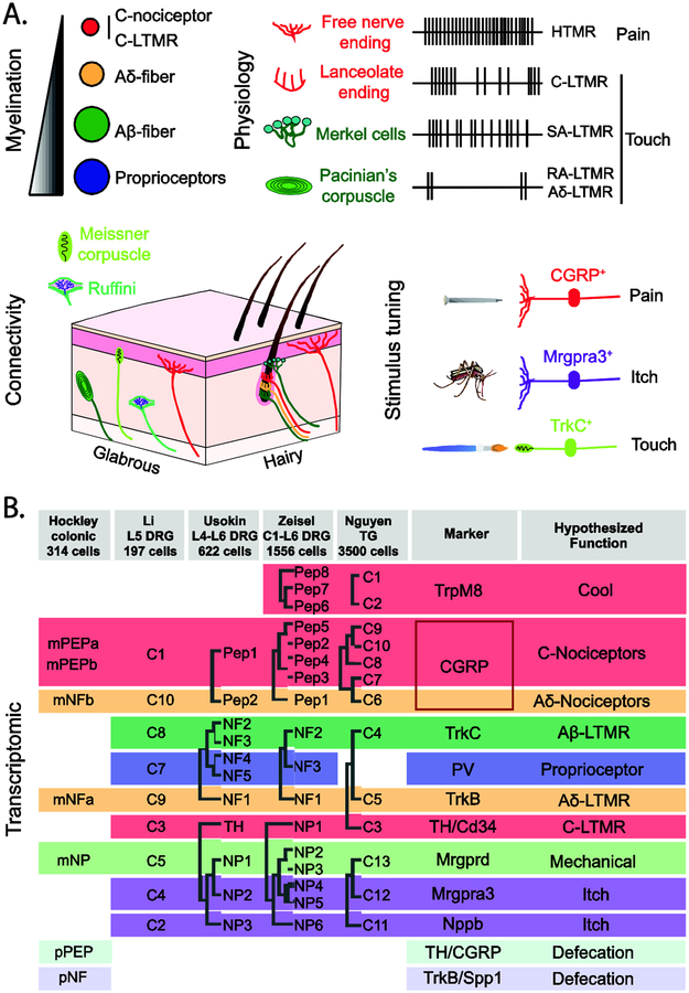
      This massive sequencing effort encompasses cells throughout the nervous system including primary afferents and dorsal horn neurons, providing an updated view of diversity: http://mousebrain.org

Publication types