. 2019 Jun;189(6):1226-1240.
doi: 10.1016/j.ajpath.2019.02.015. Epub 2019 Apr 5.

Carboxyl Ester Lipase May Not Mediate Lipotoxic Injury during Severe Acute Pancreatitis

Affiliations

Carboxyl Ester Lipase May Not Mediate Lipotoxic Injury during Severe Acute Pancreatitis

Biswajit Khatua et al. Am J Pathol. 2019 Jun.

Abstract

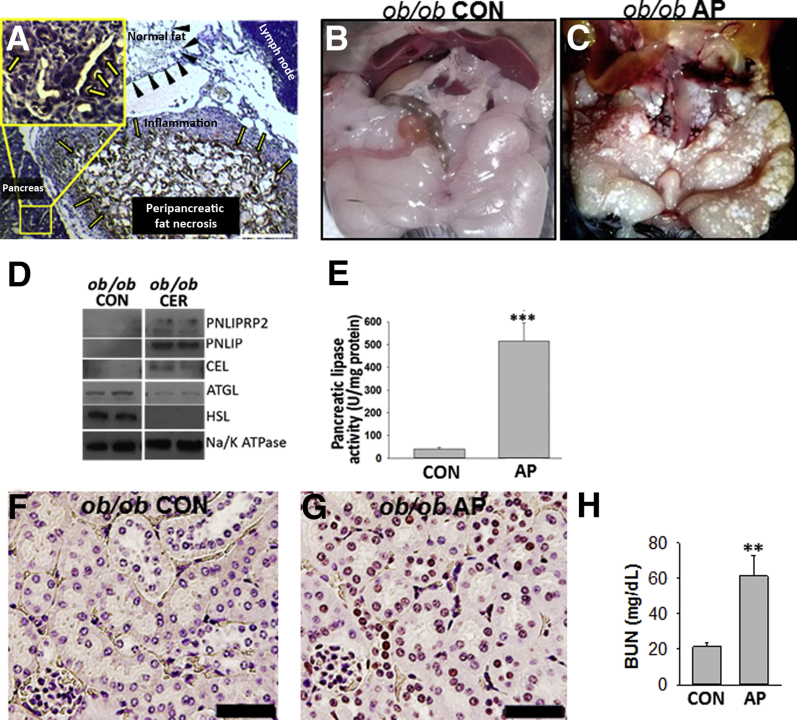
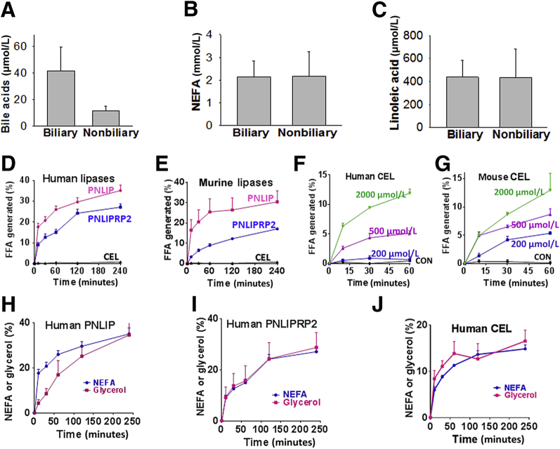
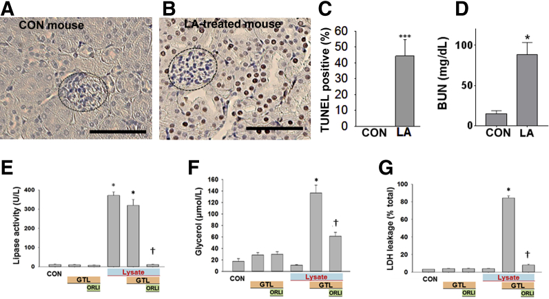
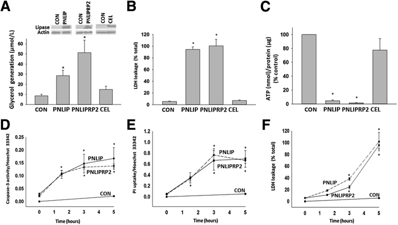
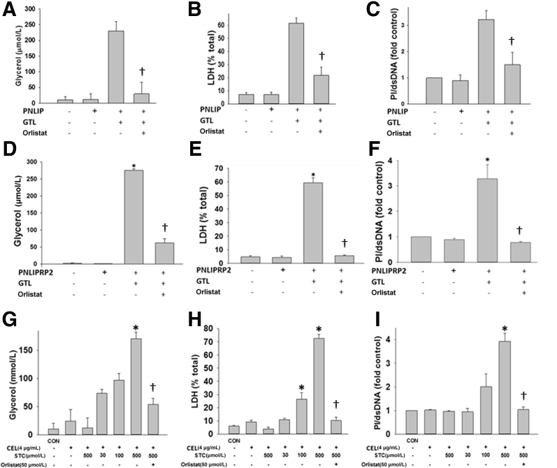
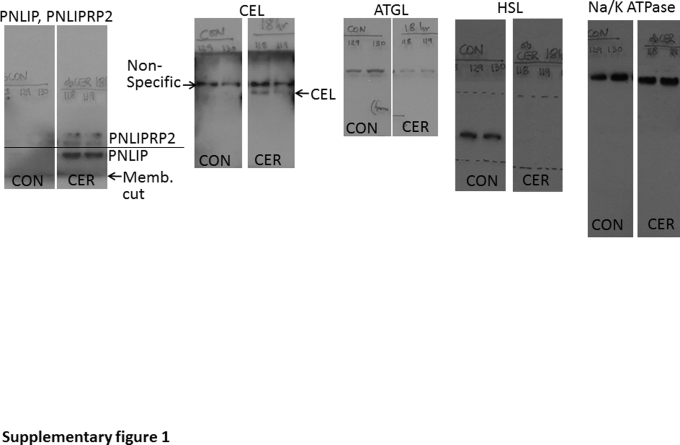
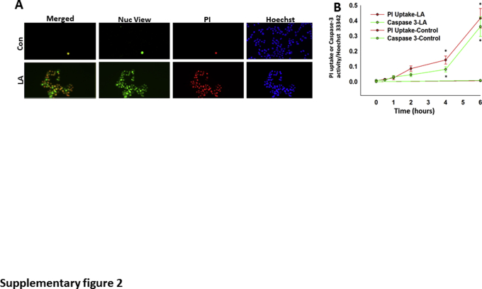
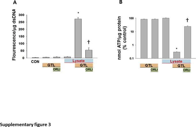
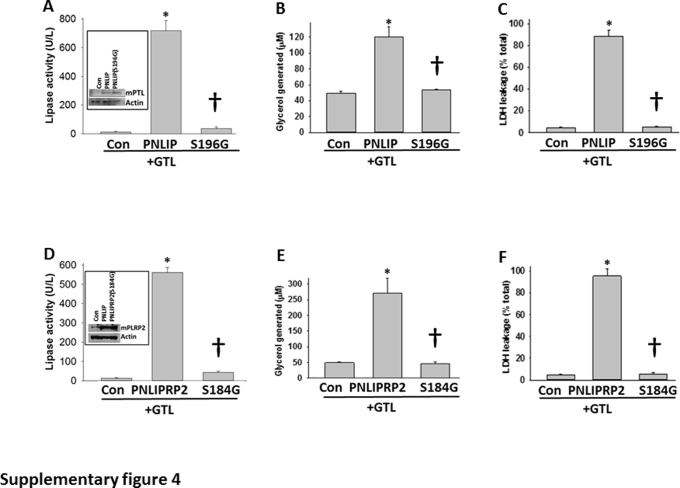
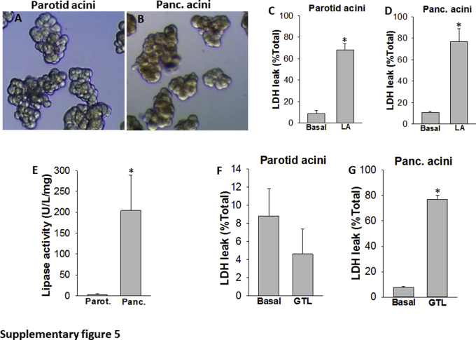
Acute lipolysis of visceral fat or circulating triglycerides may worsen acute pancreatitis (AP)-associated local and systemic injury. The pancreas expresses pancreatic triacylglycerol lipase (PNLIP), pancreatic lipase-related protein 2 (PNLIPRP2), and carboxyl ester lipase (CEL), which may leak into the visceral fat or systemic circulation during pancreatitis. We, thus, aimed to determine the pancreatic lipase(s) regulating lipotoxicity during AP. For this AP, associated fat necrosis was analyzed using Western blot analysis. Bile acid (using liquid chromatography-tandem mass spectrometry) and fatty acid (using gas chromatography) concentrations were measured in human fat necrosis. The fat necrosis milieu was simulated in vitro using glyceryl trilinoleate because linoleic acid is increased in fat necrosis. Bile acid requirements to effectively hydrolyze glyceryl trilinoleate were studied using exogenous or overexpressed lipases. The renal cell line (HEK 293) was used to study lipotoxic injury. Because dual pancreatic lipase knockouts are lethal, exocrine parotid acini lacking lipases were used to verify the results. PNLIP, PNLIPRP2, and CEL were increased in fat necrosis. Although PNLIP and PNLIPRP2 were equipotent in inducing lipolysis and lipotoxic injury, CEL required bile acid concentrations higher than in human fat necrosis. The high bile acid requirements for effective lipolysis make CEL an unlikely mediator of lipotoxic injury in AP. It remains to be explored whether PNLIP or PNLIPRP2 worsens AP severity in vivo.


Figures

Figure 1
Figure 1
Detection and activity of pancreatic lipases in fat necrosis. A: Immunohistochemistry for pancreatic triacylglycerol lipase (PNLIP) in human pancreatitis, showing positive staining in acinar cells (yellow arrows, inset) and peripancreatic fat necrosis. The inset shows the boxed area in higher magnification. Normal fat (black arrowheads) and lymph node do not stain positive. B and C: Gross images of fat pads from control (CON) ob/ob mice (note smooth glistening appearance; B) and those dying from acute pancreatitis (AP; note fat necrosis seen as numerous chalky white deposits in the fat pads; C). D: Western blot analysis of the fat pads in CON mice and in those with AP. All three pancreatic lipases are increased in AP-associated fat necrosis compared with controls. E: Bar graphs depicting pancreatic lipase activity measured in the fat pads of control mice with cerulein (CER) pancreatitis. F and G: TUNEL staining of mouse kidney in CON (F) mice and in those with AP (G). H: Blood urea nitrogen (BUN) in control mice and in those with AP. Data are expressed as means ± SEM (E and H). n = 6 to 8 mice per group (FH). ∗∗P < 0.01, ∗∗∗P < 0.001 versus control. Scale bars: 200 μm (A); 100 μm (F and G). ATGL, adipocyte triglyceride lipase; CEL, carboxyl ester lipase; HSL, hormone-sensitive lipase; PNLIPRP2, pancreatic lipase-related protein 2.
Figure 2
Figure 2
Comparison of the lipolysis by pancreatic lipases using conditions relevant to human pancreatitis. AC: Parameters [bile acid (A), nonesterified fatty acids (NEFAs; B), and linoleic acid (C)] measured in human pancreatic fluid collections of biliary and nonbiliary origin. D and E: Comparison of the ability of 1 μg/mL of human (D) and murine (E) pancreatic lipases to hydrolyze 600 μmol/L glyceryl trilinoleate (GTL) at pH 7.4 in the absence of bile acids. F and G: Effect of different concentrations of sodium taurocholate on human (F) and mouse (G) carboxyl ester lipase (CEL), as measured by the free fatty acid (FFA) generated by GTL (600 μmol/L) lipolysis over 1 hour. HJ: Comparison of the rate of FFA production with glycerol generation by GTL (600 μmol/L) lipolysis among the three pancreatic lipases. Although there is an initial delay in generating glycerol by pancreatic triacylglycerol lipase (PNLIP), by 4 hours, both glycerol and FFA are equivalent. Data are expressed as means ± SEM. CON, control; PNLIPRP2, pancreatic lipase-related protein 2.
Figure 3
Figure 3
Renal injury induced by the lipolytic product of glyceryl trilinoleate [GTL; ie, linoleic acid (LA)]. AD:In vivo renal injury induced by linoleic acid. A and B: TUNEL staining in renal tubules of control (CON) mice (A) and those given LA (B). Glomeruli (dashed ovals) were not TUNEL positive. C and D: Quantification of TUNEL-positive cells (C) and serum blood urea nitrogen (BUN) at the time of necropsy (D). EG: Effect of lipolysis of GTL (600 μmol/L) by pancreatic lysate on injury to the HEK 293 renal cell line. Lipase activity (E), glycerol (F), and lactate dehydrogenase (LDH) leakage (G), measured after the addition of pancreatic lysates (lysate) alone or in the presence of the triglyceride GTL (600 μmol/L). When used, the lipase inhibitor orlistat (ORLI; 50 μmol/L) was added immediately before addition of GTL. All these phenomena, induced in the presence of lysates and GTL, are significantly reduced by orlistat. Lipolysis of GTL is essential to induce injury. Data are expressed as means ± SEM (CG). n = 6 to 8 mice per group (AD). P < 0.05, ∗∗∗P < 0.001 versus control; P < 0.05 versus without orlistat. Scale bars: 100 μm (A and B).
Figure 4
Figure 4
Comparison of pancreatic lipase overexpression in mediating injury in the kidney cell line HEK 293. A: Mouse lipases were overexpressed in HEK 293 cells and underwent Western blot analysis (bands at top of panel) using actin as a loading control. AC: The ability to induce triglyceride hydrolysis was measured as glycerol generation (A) from the lipolysis of exogenously added glyceryl trilinoleate (GTL; 600 μmol/L) over 5 hours, and the resulting cell injury was measured as lactate dehydrogenase (LDH) leakage (B) into the medium or ATP decrease in the cell pellet (C). Note the lack of effect of carboxyl ester lipase (CEL). DF: Effect of adding GTL on the increase in active caspase-3 staining (D), propidium iodide (PI) uptake (E), and LDH leakage (F), induced by mouse pancreatic triacylglycerol lipase (PNLIP) and pancreatic lipase-related protein 2 (PNLIPRP2) versus control (CON), as a function of time. Data are expressed as means ± SEM. P < 0.05 versus control.
Figure 5
Figure 5
Bar graphs comparing human pancreatic triacylglycerol lipase (hPNLIP; 1 μg/mL), human pancreatic lipase-related protein 2 (hPNLIPRP2; 1 μg/mL), and human carboxyl ester lipase (hCEL; 4 μg/mL) on lipotoxic parotid acinar necrosis. Parotid acini were exposed to exogenous glyceryl trilinoleate (GTL; 300 μmol/L) in the presence of recombinant lipases. In case of hPNLIP and hPNLIPRP2, no bile acids were present in the medium. The concentration of sodium taurocholate (STC) used for CEL is shown under the corresponding bars. The experiments were performed for 4 hours. Lipolysis was measured by glycerol release (A, D, and G), and cell injury in terms of lactate dehydrogenase (LDH) leakage (B, E, and H) and propidium iodide (PI) uptake (C, F, and I) were measured with or without the lipase inhibitor orlistat (50 μmol/L). Note CEL required ≥100 μmol/L STC to induce cell injury. Data are expressed as means ± SEM. n = at least 3 independent experiments. P < 0.05 versus control (CON); P < 0.05 versus without orlistat. dsDNA, double-stranded DNA.
Supplemental Figure S1
Images of Western blot analyses of the fat pads of control (CON) ob/ob mice and those with 18 hours of cerulein (CER) pancreatitis, showing the film corresponding to the whole image of the membrane (Memb.) blotted for the proteins shown in Figure 1D, above and below the band of interest. The proteins for which the blot analysis was performed are mentioned at the top of the images. The antibody for pancreatic triacylglycerol lipase (PNLIP) and pancreatic lipase-related protein 2 (PNLIPRP2) detects both proteins, as described previously. ATGL, adipocyte triglyceride lipase; CEL, carboxyl ester lipase; HSL, hormone-sensitive lipase.
Supplemental Figure S2
A: Renal HEK cells [untreated control (Con; top panels)] or treated with linoleic acid (LA; 100 μmol/L; bottom panels) in serum-free media for 5 hours and stained with NucView 488 caspase-3 (Nuc), propidium iodide (PI), and Hoechst 33342. B: Quantification of the PI/Hoechst 33342 fluorescence (red lines) and NucView/Hoechst 33342 (green lines) shows a significant increase in caspase-3 activity and PI uptake with LA at 4 and 6 hours. Data are expressed as means ± SEM (B). n = 4 independents experiments (B). P < 0.05 versus control on analysis of variance.
Supplemental Figure S3
Propidium iodide uptake (A) and ATP levels (B) measured after the addition of pancreatic lysates (Lysate) alone or in the presence of the triglyceride glyceryl trilinoleate (GTL; 600 μmol/L). When used, the lipase inhibitor orlistat (ORLI; 50 μmol/L) was added immediately before addition of GTL. All these phenomena induced in the presence of lysates and GTL are significantly reduced by orlistat. Data are expressed as means ± SEM. P < 0.05 versus control (CON); P < 0.05 versus control. dsDNA, double-stranded DNA.
Supplemental Figure S4
Point mutants of mouse pancreatic triacylglycerol lipase (PNLIP) and pancreatic lipase-related protein 2 (PNLIPRP2), lacking lipase activity, do not mediate acute lipotoxicity in HEK 293 cells. A and D: Overexpression of mutant mouse Pnlip and PNLIPRP2 in HEK 293 cells, using pcDNA 3.1/PNLIP (S169G) and pcDNA 3.1/PNLIPRP2 (S184G), shows a significant decrease in pancreatic lipase activity in the medium only in the wild type (WT) and not the mutant mice, which have an equal amount of protein in the lysates (Western blot images; insets). B, C, E, and F: In the presence of glyceryl trilinoleate (GTL; 600 μmol/L), there is a significant increase in glycerol generation (B and E) and lactate dehydrogenase (LDH) leakage (C and F) in the WT, but not the mutant, mice. Data are expressed as means ± SEM. P < 0.05 versus control (Con; increase); P < 0.05 compared with the wild type (reduction).
Supplemental Figure S5
A and B: Comparison of parotid (Parot.) and pancreatic (Panc.) acini: Low-power images of parotid (A) and pancreatic (B) acini. C and D: Bar graphs comparing the lactate dehydrogenase (LDH) leakage from parotid and pancreatic acini after 4 hours of exposure to linoleic acid (LA; 300 μmol/L) with untreated (basal) acini. E: Pancreatic lipase activity/mg protein in the homogenates of mouse parotid and pancreas. F and G: LDH leakage from parotid (F) and pancreatic (G) acini after 4 hours of incubation under the basal state or with 300 μmol/L glyceryl trilinoleate (GTL). Data are expressed as means ± SEM. P < 0.05 versus control. Original magnification, ×10 (A and B).

References

    1. Navina S., Acharya C., DeLany J.P., Orlichenko L.S., Baty C.J., Shiva S.S., Durgampudi C., Karlsson J.M., Lee K., Bae K.T., Furlan A., Behari J., Liu S., McHale T., Nichols L., Papachristou G.I., Yadav D., Singh V.P. Lipotoxicity causes multisystem organ failure and exacerbates acute pancreatitis in obesity. Sci Transl Med. 2011;3:107ra10. - PMC - PubMed
    1. Durgampudi C., Noel P., Patel K., Cline R., Trivedi R.N., DeLany J.P., Yadav D., Papachristou G.I., Lee K., Acharya C., Jaligama D., Navina S., Murad F., Singh V.P. Acute lipotoxicity regulates severity of biliary acute pancreatitis without affecting its initiation. Am J Pathol. 2014;184:1773–1784. - PMC - PubMed
    1. Noel P., Patel K., Durgampudi C., Trivedi R.N., de Oliveira C., Crowell M.D., Pannala R., Lee K., Brand R., Chennat J., Slivka A., Papachristou G.I., Khalid A., Whitcomb D.C., DeLany J.P., Cline R.A., Acharya C., Jaligama D., Murad F.M., Yadav D., Navina S., Singh V.P. Peripancreatic fat necrosis worsens acute pancreatitis independent of pancreatic necrosis via unsaturated fatty acids increased in human pancreatic necrosis collections. Gut. 2016;65:100–111. - PMC - PubMed
    1. Patel K., Trivedi R.N., Durgampudi C., Noel P., Cline R.A., DeLany J.P., Navina S., Singh V.P. Lipolysis of visceral adipocyte triglyceride by pancreatic lipases converts mild acute pancreatitis to severe pancreatitis independent of necrosis and inflammation. Am J Pathol. 2015;185:808–819. - PMC - PubMed
    1. Nawaz H., Koutroumpakis E., Easler J., Slivka A., Whitcomb D.C., Singh V.P., Yadav D., Papachristou G.I. Elevated serum triglycerides are independently associated with persistent organ failure in acute pancreatitis. Am J Gastroenterol. 2015;110:1497–1503. - PubMed

Publication types