. 2019 Jun:190:183-192.
doi: 10.1016/j.jsbmb.2019.04.003. Epub 2019 Apr 4.

Sp1 regulates steroidogenic genes and LHCGR expression in primary human luteinized granulosa cells

Affiliations

Sp1 regulates steroidogenic genes and LHCGR expression in primary human luteinized granulosa cells

Scott Convissar et al. J Steroid Biochem Mol Biol. 2019 Jun.

Abstract

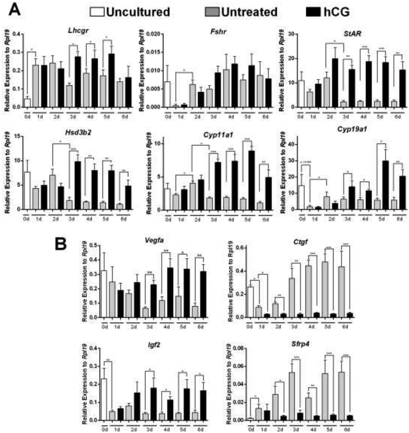
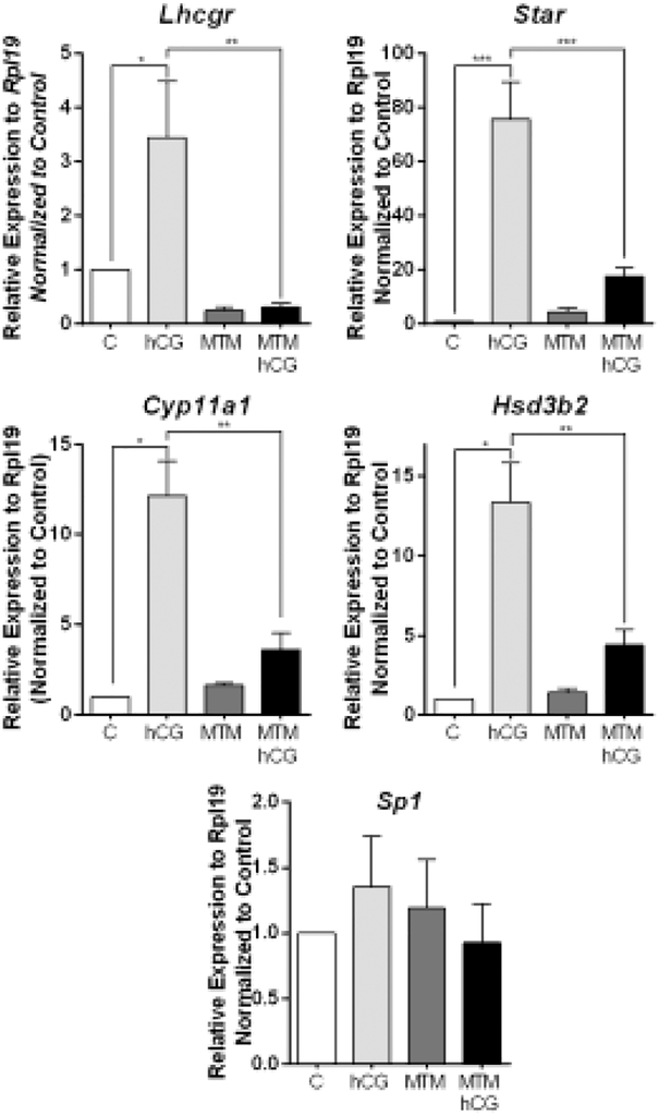
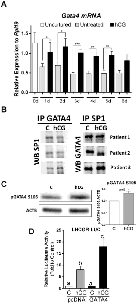
Luteinizing hormone and human chorionic gonadotropin (hCG) bind to the luteinizing hormone/chorionic gonadotropin receptor (LHCGR). LHCGR is required to maintain corpus luteum function but the mechanisms involved in the regulation of LHCGR in human luteal cells remain incompletely understood. This study aimed to characterize the expression of LHCGR mRNA in primary human luteinized granulosa cells (hLGCs) obtained from patients undergoing in vitro fertilization and to correlate LHCGR expression with the response of hLGCs to hCG by assessing the expression of genes known to be markers of hCG actions. The results show that LHCGR expression is low in freshly isolated cells but recovers rapidly in culture and that hCG maintains LHCGR expression, suggesting a positive feedback loop. The activity of a LHCGR-LUC reporter increased in cells treated with hCG but not with follicle-stimulating hormone. Treatment with hCG also stimulated the expression of genes involved in steroidogenesis in a time-dependent manner. LHCGR promoter expression was found to be regulated by SP1, which we show is highly expressed in hLGCs. Moreover, SP1 inhibition prevented the stimulation of steroidogenic genes and the increase in LHCGR-LUC reporter activity by hCG. Finally, we provide evidence that a complex formed by SP1 and GATA4 may play a role in the maintenance of LHCGR expression. This report reveals the mechanisms involved in the regulation of the LHCGR and provides experimental data demonstrating that the proximal region of the LHCGR promoter is sufficient to drive the expression of this gene in primary hLGCs.

Keywords: GATA factors; Human luteal cells; Luteinizing hormone; Progesterone production; SP1; Steroidogenesis.


Conflict of interest statement

Declaration of interest

The authors declare that there is no conflict of interest that could be perceived as prejudicing the impartiality of the research reported.

Figures

Figure 1:
Figure 1:. Time-course of gene expression in the presence or absence of hCG.
Human LGCs were obtained after ovarian hyperstimulation from IVF patients at the time of follicle aspiration. Cells were harvested immediately (Uncultured, 0d), or cultured in serum-free media for a period of 1, 2, 3, 4, 5, or 6 days in the presence or absence of recombinant hCG (50 ng/ul). Gene expression was quantified using qPCR. Figure 1A represents LHCGR, FSHR and steroidogenic gene expression, while Figure 1B represents non-steroidogenic gene expression (see Results section). Columns represent the mean ± SEM of six different samples. *P < 0.05, **P < 0.01, ***P < 0.001 vs. C, t-test n=6.
Figure 2:
Figure 2:. Effect of gonadotropins on LHCGR promoter activity in primary hLGCs.
Human LGCs were plated for 48 h in serum-free media followed by a 48-hour incubation period with a lentiviral vector carrying a 174 bp region of the human LHCGR proximal promoter. At this point, the cells were either treated with vehicle (C), FSH (50 ng/ml), or hCG (50 ng/ml) for 6, 24, or 48 h and harvested for luciferase reporter assay. *P > 0.05, t-test against specific time-point control, n=6.
Figure 3:
Figure 3:
A) LHCGR promoter - Comparison of human (−174 bp), bovine (−167 bp), and murine (−164 bp) LHCGR promoter regions showing a high degree of conservation between the species or the SP1 sites. B) SP1 mRNA expression in cultured hLGCs. Sp1 mRNA levels were quantified in hLGCs treated as in Figure 1 by qPCR (n=6). C) hLGCs from 3 patients were plated separately and cultured for 48 h in serum-free media to recover LHCGR expression followed by a 48-hour treatment with hCG. Protein expression was assessed by western blot for SP1 (106 kDa and 95 kDa bands) and ACTB as a loading control. Densiometric analysis of protein signal is also shown (ns=not significant, n=3).
Figure 4:
Figure 4:. Mutation of SP1 sites blunts the activation of the LHCGR promoter.
hLGCs were plated for 24 h in serum-free media followed by a 48-hour incubation with a lentiviral vector carrying a control reporter plasmid, a plasmid containing an intact proximal human LHCGR promoter, or a plasmid containing mutations in the SP1 sites of the LHCGR promoter. Cells were harvested after no treatment (C) or treatment with hCG (50 ng/ml), forskolin (5 μM), or dbcAMP (1 mM) for 48 h. Quantitation of promoter activity was performed using a luciferase reporter assay. *P < 0.05 t-test, n = 6.
Figure 5:
Figure 5:. Effect of LHCGR pathway inhibition on human LHCGR promoter activity.
hLGCs were cultured for 24 h in serum-free media then transfected with the 174 bp human LHCGR proximal promoter region. Cells were left untreated (C) or were treated with hCG (50 ng/ml) or forskolin (5 μM) for 48 h in the presence or absence of H89 (H), U0126 (U), MK2206 (MK) or Mithramycin A (MTM). Paired columns differ significantly from each other by t-test (P < 0.05, n = 6).
Figure 6:
Figure 6:. Inhibition of SP1 blocks the response of hLGCs to hCG.
Primary hLGCs were plated for 48 h in serum-free media followed by 48 h incubation with hCG, MTM, or hCG+MTM. Quantification of the expression of Lhcgr, Star, Cyp11a1, Hsd3b2, and Sp1 was performed using qPCR. Columns represent the mean ± SEM of six different samples. (*P < 0.05, **P < 0.01, ***P < 0.001 vs. C, one-way ANOVA, Tukey test).
Figure 7:
Figure 7:. GATA4 may play a role in the regulation of the proximal promoter of the LHCGR gene.
A) Cells were treated as indicated in Figure 1. The expression of Gata4 was examined by qPCR. Columns represent the mean ± SEM of six different samples. *P < 0.05, **P < 0.01, ***P < 0.001 vs. C, t-test n=6. B) Cells from three patients were cultured in separate 6-well plates for 48 h and then treated with vehicle or hCG for 1 hour. GATA4 and SP1 were immunoprecipitated. Then western blot was used to detect GATA4 in SP1 precipitates or SP1 in GATA4 precipitates. C) Cells were treated with vehicle or hCG for 1 hour. Western blot for phospho-GATA4 and β-actin (BACT) was performed. The experiment was repeated with 5 different patients, a representative blot is shown, and results quantified and expressed as the ratio of pGATA4/BACT. Columns represent the mean ± SEM, *P < 0.05 vs. C, t-Test, n=5. D) Cells were infected with virus carrying LHCGR reporter and virus carrying an empty cassette or a GATA4 expression cDNA. 48 h later, cells were treated with vehicle or hCG for 48 h. Columns represent the mean ± SEM of six different experiments. Columns with different letters differ significantly (P < 0.05; one-way ANOVA, Tukey test).

References

    1. Stocco C, Telleria C, Gibori G, The molecular control of corpus luteum formation, function, and regression, Endocr Rev, 28 (2007) 117–149. - PubMed
    1. Lei ZM, Mishra S, Zou W, Xu B, Foltz M, Li X, Rao CV, Targeted disruption of luteinizing hormone/human chorionic gonadotropin receptor gene, Mol Endocrinol, 15 (2001) 184–200. - PubMed
    1. Zhang FP, Poutanen M, Wilbertz J, Huhtaniemi I, Normal prenatal but arrested postnatal sexual development of luteinizing hormone receptor knockout (LuRKO) mice, Mol Endocrinol, 15 (2001) 172–183. - PubMed
    1. Hamalainen T, Poutanen M, Huhtaniemi I, Promoter function of different lengths of the murine luteinizing hormone receptor gene 5’-flanking region in transfected gonadal cells and in transgenic mice, Endocrinology, 142 (2001) 2427–2434. - PubMed
    1. Nakao K, Kishi H, Imai F, Suwa H, Hirakawa T, Minegishi T, TNF-alpha suppressed FSH-induced LH receptor expression through transcriptional regulation in rat granulosa cells, Endocrinology, 156 (2015) 3192–3202. - PubMed

Publication types

LinkOut - more resources