Review
. 2019 Jun:56:175-184.
doi: 10.1016/j.conb.2019.03.003. Epub 2019 Apr 5.

Cell type and circuit modules in the spinal cord

Affiliations
Review

Cell type and circuit modules in the spinal cord

Peter J Osseward 2nd et al. Curr Opin Neurobiol. 2019 Jun.

Abstract

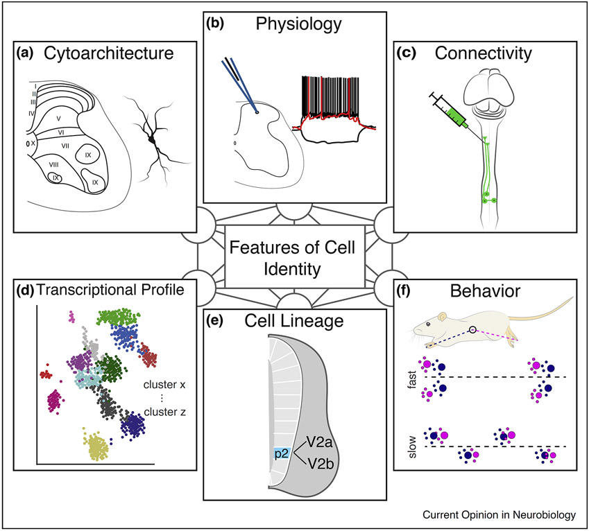
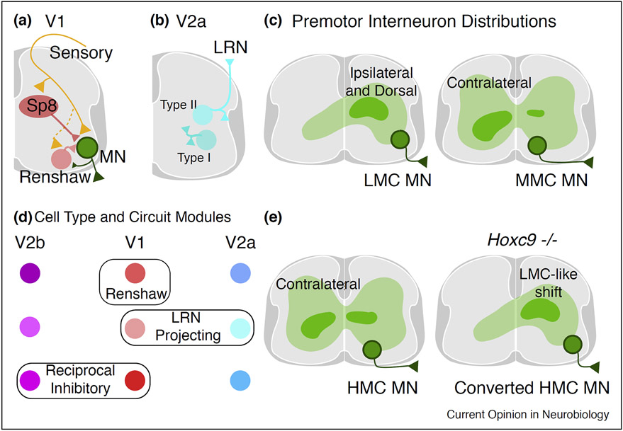
The spinal cord contains an extraordinarily diverse population of interconnected neurons to process somatosensory information and execute movement. Studies of the embryonic spinal cord have elucidated basic principles underlying the specification of spinal cord neurons, while adult and postnatal studies have provided insight into cell type function and circuitry. However, the overarching principles that bridge molecularly defined subtypes with their connectivity, physiology, and function remain unclear. This review consolidates recent work in spinal neuron characterization, examining how molecular and spatial features of individual spinal neuron types relate to the reference points of connectivity and function. This review will focus on how spinal neuron subtypes are organized to control movement in the mouse.


Conflict of interest statement

Conflict of interest statement

Nothing declared.

Figures

Figure 1
Figure 1. Features encompassing neuronal identity in the spinal cord. Spinal neurons can be classified based on many individual features, but the relationships between these features are still unclear.
(a) Spinal neurons can be classified based on cell morphology and location. Laminar designation, originally described by Bror Rexed, is a common method to categorize spinal neurons. (b) Spinal neurons possess a variety of intrinsic electrophysiological properties. Furthermore, cell recordings paired with stimulation (such as dorsal root stimulation) can reveal functional connections between neuronal populations. (c) Various tracers and viral approaches have been used to examine connectivity between spinal neurons. Anterograde, retrograde, and trans-synaptic tracing have been used to identify neuronal populations with specific projection patterns. (d) RNA sequencing, both at the population and single cell level, have identified many transcriptionally distinct populations within the spinal cord. (e) Developmental studies have examined genes involved in the specification of spinal neurons. Overexpression and knockout studies have provided insight into gene function, while lineage tracing has allowed investigators to probe the anatomy and function of developmentally defined cell types. (f) Spinal neurons can be classified based on their role in behaviors. Early activity gene markers such as c-fos have been used to identify cohorts of neurons active during specific behaviors. Activation, silencing, and ablation studies have allowed spinal neurons to be categorized by the behaviors they mediate.
Figure 2
Figure 2. Spinal cord regions and cell types.
(a) Spinal regions and corresponding Rexed laminae. (b) Examples of molecularly defined cell types located in each region. In some cases, the gene of interest is also expressed in other spinal regions. Example dorsal cell types are RORα, GRP, and NPY expressing neurons [4,9,52-54]. Ventral cell types include En1, Chx10, and Hb9 expressing neurons [2,32••,34••]. Gad2 and Satb2 expressing neurons are intermediate spinal cord cell types [19,22,26-28]. (c) Exteroceptive sensory fibers terminate in the dorsal cord in a topographic fashion [4,7,8]. Noxious sensory afferents primarily terminate within laminae I–II. Low threshold mechanoreceptors terminate within laminae II–IV of the spinal cord. (d) Motor neurons are organized into columns and pools which reflect a musculotopic map [10-12]. Lumbar ventral interneurons form a network named the locomotor central pattern generator (CPG), which rhythmically excites motor neurons to generate locomotion [3,13]. (e) Many intermediate interneurons are monosynaptically connected to motor neurons [19,21]. Stimulation of intermediate premotor neurons evokes synergistic activity in multiple motor pools, thus these neurons are designated motor synergy encoders (MSE) [19]. The intermediate spinal cord receives a variety of sensory and descending inputs. Dorsal interneurons relay exteroceptive information onto intermediate spinal interneurons [20]. The corticospinal tract and proprioceptive afferents also densely terminate in the intermediate spinal cord [19,21]. Proprioceptive afferents also innervate motor neurons and subsets of ventral interneurons.
Figure 3
Figure 3. Molecular organization of spinal neurons.
(a) During embryonic development, spinal interneurons are derived from 13 progenitor domains (including the late born dILa and dILb domains). Highlighted are the pMN, p1, and p2 domains. After neurogenesis, cells migrate to their final settling positions [2]. (b) The p1 domain generates V1 interneurons, which express En1. V1 interneurons settle throughout the ventral horn. The majority of V1 interneurons can be divided into 4 clades, expressing either Sp8, Pou6f2, FoxP2, or MafA. V1 clades can be further divided into many subtypes based on combinatorial expression of additional transcription factors. These additional transcriptional divisions often correspond to spatial divisions as well [32••]. (c) The p2 domain generates V2a–V2d interneurons [2,55]. V2a interneurons are divided into a medial and lateral column based on Nfib and Zfhx3 expression. Each column comprises multiple transcriptionally distinct subtypes; however, the spatial locations of these subtypes have yet to be examined in detail [34••]. (d) The pMN domain gives rise to motor neurons, which express Hb9 and settle in the ventral spinal cord. Motor neurons are organized into columns. While all motor neurons initially express Lhx3, only the MMC (Medial motor column) maintains its expression. The LMC (Lateral motor column) expresses FoxP1. Within each column, motor neurons can be further divided into motor pools, each of which is spatially clustered and has a unique muscle target [10-12]. (e–g) Categories of neurons based on their rostrocaudal distribution. Constant: a population of neurons which are present throughout the length of the spinal cord. Limb: neurons which reside in the cervical and lumbar cord. Thoracic: neurons which are located in the thoracic cord. Gradient: neurons that are arrayed in a gradient along the cord. (e) V1 interneuron clades are preserved across the rostrocaudal axis. However, V1 subtypes specific to limb segments or thoracic segments can be identified based on the expression of two transcription factors [38••]. (f) V2a columns span the rostrocaudal axis. The composition of each column is a combination of Type I V2a interneurons (which maintain Chx10 expression) and Type II V2a interneurons (which downregulate Chx10). The ratio of the two types remains constant in the medial column. In the lateral column, the ratio is organized in a gradient manner which is dependent on spinal segment. Cervical segments are composed of primarily Type II V2a interneurons, while lumbar segments are primarily Type I V2a interneurons [34••]. (g) The MMC, HMC (Hypaxial motor column), and LMC synapse onto axial, intercostal, and limb muscles, respectively. The MMC spans the spinal cord, while the HMC and LMC are segment specific. Motor pools are segmentally organized as well [10-12]. Example LMC motor pools are shown (MMC and HMC pools are not shown). Motor pools generally span a few segments [10,12].
Figure 4
Figure 4. Specificity in spinal cord circuitry.
(a) Microcircuits in V1 interneurons for the Sp8 clade and Renshaw cells. Sp8 neurons receive proprioceptive input from Gluteus (GL) and Tibialis Anterior (TA) muscles while Renshaw cells only receive input from the GL. Renshaw cells receive input from GL and TA motor neurons while Sp8 neurons do not. Both Sp8 neurons and Renshaw cells synapse onto GL, TA, and intrinsic foot (IF) motor neurons [32••]. (b) Type II V2a interneurons, which downregulate Chx10 during development, are predominately located in cervical segments and project supraspinally to the lateral reticular nucleus (LRN). Type I V2a interneurons, which maintain Chx10 expression, project within the spinal cord [34••]. (c) Spatial distribution of premotor interneurons for different motor neuron column types. LMC (Lateral motor column) neurons predominately receive input from spinal interneurons located ipsilaterally and dorsally. MMC (Medial motor column) neurons receive a greater portion of input from contralaterally located spinal interneurons [19,21,42]. (d) Schematic depicting the relationship between spinal interneuron classes and circuit subtypes. Renshaw cells, which provide feedback inhibition to motor neurons, are a subtype exclusively found in V1 interneurons [49-51]. Subtypes of both V1 and V2a interneurons project to the brainstem lateral reticular nucleus (LRN) [39]. Reciprocal inhibitory neurons receive proprioceptive input from a limb muscle and inhibit the corresponding antagonist motor pool. Subtypes of V1 and V2b interneurons are reciprocal inhibitory neurons [51]. (e) Location of premotor HMC interneurons in the thoracic spinal cord. In Hoxc9 mutants, HMC neurons are converted into an LMC fate and their premotor interneuron distribution shifts accordingly [41••,42].

References

    1. Alaynick WA, Jessell TM, Pfaff SL: Snapshot: spinal cord development. Cell 2011, 146 178–178 e171. - PMC - PubMed
    1. Lu DC, Niu T, Alaynick WA: Molecular and cellular development of spinal cord locomotor circuitry. Front Mol Neurosci 2015, 8:25. - PMC - PubMed
    1. Kiehn O: Decoding the organization of spinal circuits that control locomotion. Nat Rev Neurosci 2016, 17:224–238. - PMC - PubMed
    1. Koch SC, Acton D, Goulding M: Spinal circuits for touch, pain, and itch. Annu Rev Physiol 2018, 80:189–217. - PMC - PubMed
    1. Gosgnach S, Bikoff JB, Dougherty KJ, El Manira A, Lanuza GM, Zhang Y: Delineating the diversity of spinal interneurons in locomotor circuits. J Neurosci 2017, 37:10835–10841. - PMC - PubMed

Publication types