

Early Detection of Multiorgan Light-Chain Amyloidosis by Whole-Body 18F-Florbetapir PET/CT
- PMID: 30954943
- PMCID: PMC6735282
- DOI: 10.2967/jnumed.118.221770
Early Detection of Multiorgan Light-Chain Amyloidosis by Whole-Body 18F-Florbetapir PET/CT
Abstract
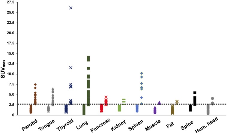
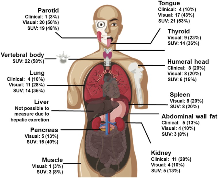
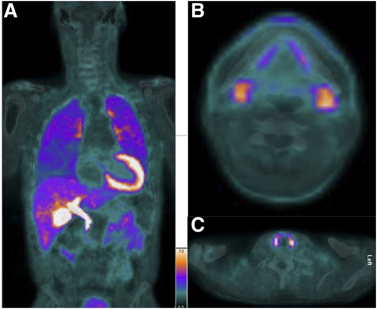
Immunoglobulin light-chain (AL) amyloidosis affects multiple systemic organs. However, determination of the precise extent of organ involvement remains challenging. Targeted amyloid imaging with 18F-florbetapir PET/CT offers the potential to detect AL deposits in multiple organs. The primary aim of this study was to determine the distribution and frequency of AL deposits in the various organs of subjects with systemic AL amyloidosis using 18F-florbetapir PET/CT. Methods: This prospective study included 40 subjects with biopsy-proven AL amyloidosis including active AL amyloidosis (n = 30) or AL amyloidosis in hematologic remission for more than 1 y (n = 10). All subjects underwent 18F-florbetapir PET/CT, skull base to below the kidney scan field, from 60 to 90 min after injection of radiotracer. Volume-of-interest measurements of SUVmax were obtained using Hermes software for the parotid gland, tongue, thyroid, lung, gastric wall, pancreas, spleen, kidney, muscle, abdominal fat, lower thoracic spine, vertebral body, and humeral head. Uptake in each organ was visually compared with that in spine bone marrow. An SUVmax of at least 2.5 was considered abnormal in all organs other than the liver. Results: Compared with the international consensus definition of organ involvement, 18F-florbetapir PET/CT identified amyloid deposits in substantially higher percentages of subjects for several organ systems, including parotid gland (50% vs. 3%), tongue (53% vs. 10%), and lung (35% vs. 10%). In several organ systems, including kidney (13% vs. 28%) and abdominal wall fat (10% vs. 13%), PET identified involvement in fewer subjects than did international consensus. Quantitative analysis of 18F-florbetapir PET/CT revealed more frequent organ involvement than did visual analysis in the tongue, thyroid, lung, pancreas, kidney, muscle, and humeral head. Extensive organ amyloid deposits were observed in active AL as well as in AL remission cohorts, and in both cardiac and noncardiac AL cohorts. Conclusion:18F-florbetapir PET/CT detected widespread organ amyloid deposition in subjects with both active AL and AL hematologic remission. In most instances, amyloid deposits in the various organs were not associated with clinical symptoms and, thus, were unrecognized. Early recognition of systemic organ involvement may help tailor treatment, and noninvasive monitoring of organ-level disease may guide management with novel fibril-resorbing therapies.
Keywords: 18F-florbetapir; AL; PET/CT; organ; systemic light chain amyloidosis.
© 2019 by the Society of Nuclear Medicine and Molecular Imaging.
Figures
References
-
- Bellotti V, Nuvolone M, Giorgetti S, et al. The workings of the amyloid diseases. Ann Med. 2007;39:200–207. - PubMed
-
- Glenner GG. Amyloid deposits and amyloidosis: the beta-fibrilloses (first of two parts). N Engl J Med. 1980;302:1283–1292. - PubMed
-
- Merlini G, Comenzo RL, Seldin DC, Wechalekar A, Gertz MA. Immunoglobulin light chain amyloidosis. Expert Rev Hematol. 2014;7:143–156. - PubMed
-
- Kyle RA, Gertz MA. Primary systemic amyloidosis: clinical and laboratory features in 474 cases. Semin Hematol. 1995;32:45–59. - PubMed
Publication types
MeSH terms
Substances
Grants and funding
LinkOut - more resources
Full Text Sources
Medical