. 2019 Mar-Apr;19(2):105-111.
doi: 10.1177/1535759719835355.

Current Controversy: Spikes, Bursts, and Synchrony in Generalized Absence Epilepsy: Unresolved Questions Regarding Thalamocortical Synchrony in Absence Epilepsy

Affiliations

Current Controversy: Spikes, Bursts, and Synchrony in Generalized Absence Epilepsy: Unresolved Questions Regarding Thalamocortical Synchrony in Absence Epilepsy

John Huguenard. Epilepsy Curr. 2019 Mar-Apr.

Abstract

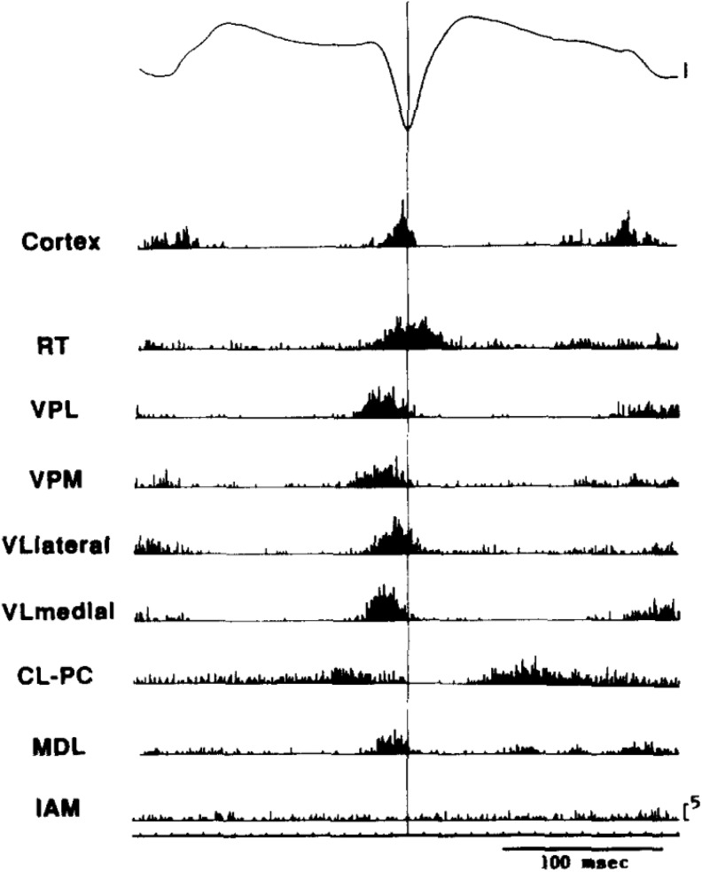
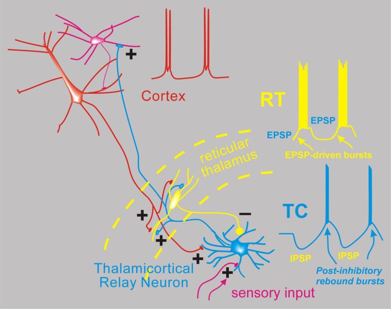
Absence epilepsy is a disorder of thalamocortical networks. Animal models have provided detailed information regarding the core cellular, synaptic, and network features that contribute to the electroencephalogram spike and wave discharge characteristic of typical absence epilepsy. Understanding of seizure networks and dynamics is a critical step toward improving treatments, yet competing conceptual models have evolved to explain seizure initiation and propagation. Recent studies have questioned 2 key model concepts: (1) T-type Ca2+ channel-dependent burst firing in thalamic relay neurons may not be essential for seizure generation, bringing into question the proposed mechanism for the antiepileptic drug ethosuximide in reducing thalamic bursting and (2) widespread synchronized neural activity may not be a core feature of the seizures, indicating that reductions in synchrony would not be a productive therapeutic goal. In this review, I will discuss these current findings, highlight the innovative approaches that have enabled these insights, and provide a unified framework that incorporates these sometimes-conflicting ideas. Finally, I lay out future work that will be necessary to finally resolve the remaining issues.


Conflict of interest statement

Declaration of Conflicting Interests: The author(s) declared no potential conflicts of interest with respect to the research, authorship, and/or publication of this article.

Figures

Figure 1.
Figure 1.
Widespread synchrony and participation of thalamic and cortical neurons during spike wave discharges in the WAG/Rij genetic rat model of absence seizures. Signals were aligned and averaged across many individual spike wave cycles. Multiunit spikes were detected in multiple electrode locations throughout the thalamus. Note that coordinated spiking occurs in all cortical and thalamic locations (except IAM and perhaps CL-PC) and that peak spiking rate occurs very near the peak of the EEG spike, with very little spiking outside this time frame, suggesting synchronous firing and coordinated pauses across the thalamocortical axis. Adapted with permission from Inoue et al. CL-PC indicates centrolateral–paracentral; EEG, electroencephalogram; IAM, interanteromedial; MDL, mediodorsal, lateral portion; RT, reticular thalamus; VPL, ventroposteriolateral; VPM, ventroposterior medial; VLlateral, ventrolateral, lateral portion; VLlmedial, ventrolateral medial portion.
Figure 2.
Figure 2.
Simplified network model for absence seizures. Depicted are interconnected neurons of the thalamus and cortex and the spiking activity proposed to occur during spike wave discharge of an absence seizure. From the bottom, thalamocortical (TC) relay neurons (blue) normally receive sensory input in the form of excitatory synapses (+) and transform this into TC cell spikes that propagate the sensory signal to the cortex (red). Thalamocortical neurons emit axon collaterals into the reticular thalamus (RT, yellow), where excitatory synapses activate the resident inhibitory neurons. These then provide feedback inhibition (inhibitory post-synaptic potentials [IPSPs]) to TC cells. During absence seizures, the network presumably becomes synchronized. In this scenario, multiple RT cells fire together producing strong IPSPs onto TC cells, and since TC cells contain high levels of T type calcium channels, strong inhibition leads to robust postinhibitory rebound bursts that in turn reactivate both RT and cortex (excitatory blue axonal fibers). Cortical cell firing (red) is also synchronized so that its excitatory output can combine with TC output to strongly activate RT cells, which also contain T-type calcium channels and generate bursts of action potentials via direct synaptic excitation. In this model, cells from all 3 classes (Cortex, TC, RT) actively participate in a near synchronous manner, with the inhibitory output from RT delaying TC cell bursting and thus playing a major role in pacing the epileptic network.

References

    1. Hunter J, Jasper H. Reactions of unanaesthetised animals to thalamic stimulation. Trans Am Neurol Assoc. 1948;73(73 Annual Meet):171. - PubMed
    1. Gloor P, Avoli M, Kostopoulos G. Thalamo-cortical relationships in generalized epilepsy with bilaterally synchronous spike-and-wave discharge In Generalized Epilepsy: Neurobiological Approaches, Boston, MA: Birkhäuser; 1990;190–212.
    1. Fogerson PM, Huguenard JR. Tapping the brakes: cellular and synaptic mechanisms that regulate thalamic oscillations. Neuron. 2016;92(4):687–704. - PMC - PubMed
    1. Maheshwari A, Noebels JL. Monogenic models of absence epilepsy: windows into the complex balance between inhibition and excitation in thalamocortical microcircuits. Prog Brain Res. 2014;213:223–252. - PubMed
    1. McCormick DA, Contreras D. On the cellular and network bases of epileptic seizures. Annu Rev Physiol. 2001;63:815–846. - PubMed

LinkOut - more resources