

Non-squamous cell carcinoma diseases of the larynx: clinical and imaging findings
- PMID: 30956151
- PMCID: PMC9422427
- DOI: 10.1016/j.bjorl.2019.02.003
Non-squamous cell carcinoma diseases of the larynx: clinical and imaging findings
Abstract
Introduction: Squamous cell carcinoma is the most common laryngeal neoplasm and accounts for approximately 95% of all malignant neoplams of the larynx. However, various benign and malignant tumors and inflammatory diseases may affect the larynx.
Objective: The purpose of this study is to analyze the clinical and imaging findings of non-squamous cell neoplasms and inflammatory diseases of the larynx.
Methods: This retrospective study was conducted in 18 patients who were diagnosed with non-squamous cell carcinoma lesions of larynx at our institution between 2007-2017. Clinical symptoms, examination findings, imaging characteristics, histopathologic diagnosis and treatment modalities were analyzed.
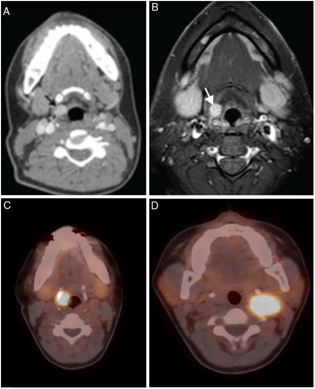
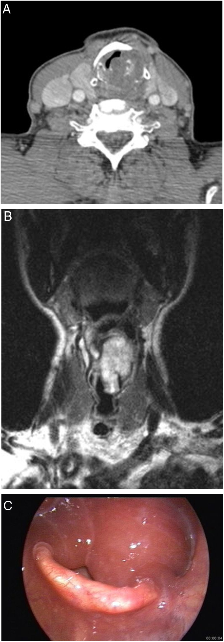
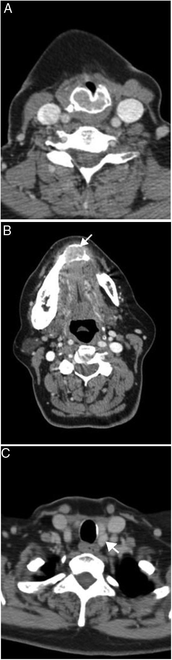
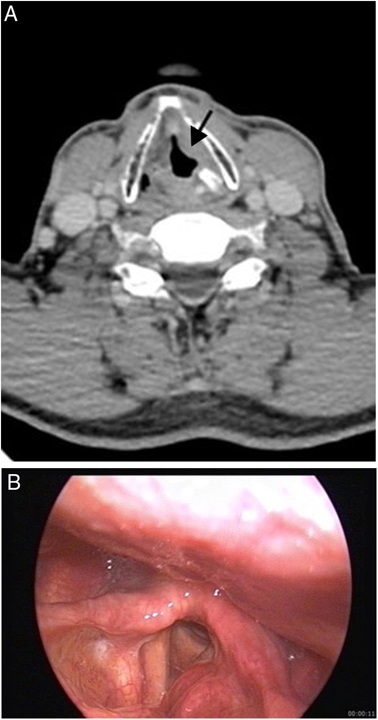
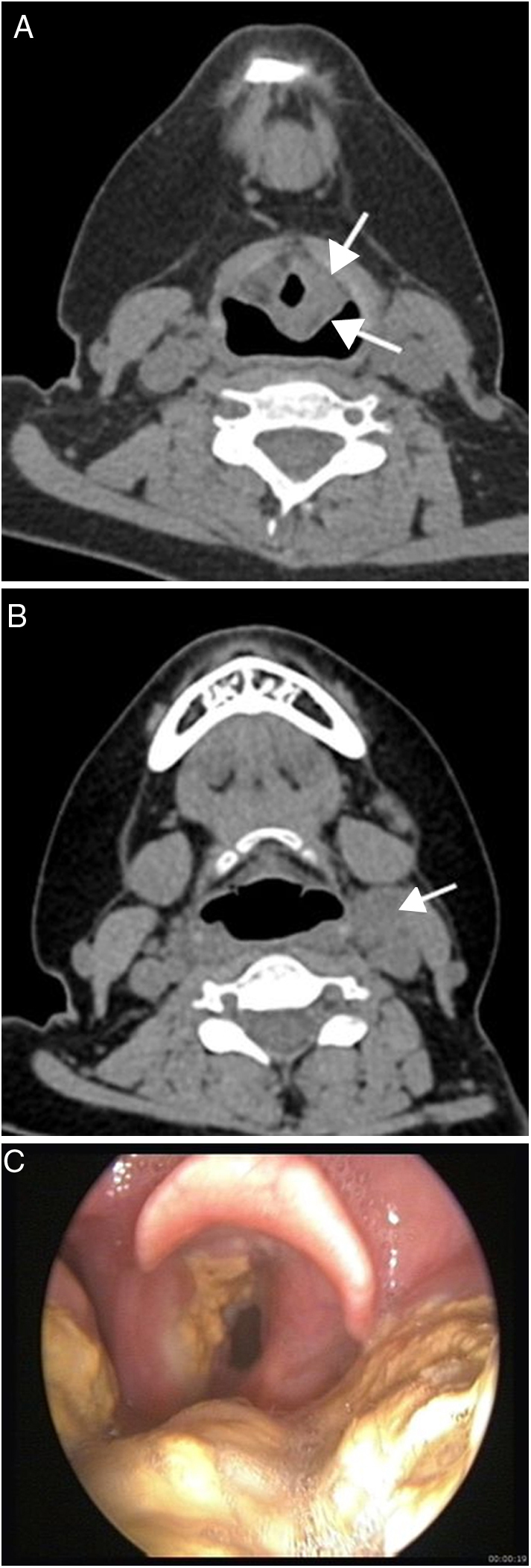
Results: There were 9 malignant lesions (2 chondrosarcoma, 1 neuroendocrine tumor-atipical carcinoid, 1 Natural Killer/T-cell lymphoma, 1 diffuse large B-cell lymphoma, 3 plasmocytoma-multiple myeloma involvement, 1 adenocarcinoma metastasis), 3 benign neoplasms (chondroma, paraganglioma, lipoma), 2 tumor-like lesions (Brown tumor and inflammatory myofibroblastic tumor), 3 inflammatory lesions (Wegener granulomatosis, Behçet's disease and tuberculosis involvements), and 1 vascular malformation. The most common presenting symptom was hoarseness (66.6%). Paraganglioma was seen as hypervascular lesion on computed tomography and magnetic resonance imaging and showed intense tracer uptake on 68Gallium-DOTA-peptide PET/CT. Chondroid matrix calcifications were detected in chondroma and chondrosarcoma-grade 1. In patients with vascular malformation and lipoma, the typical imaging findings made it possible to diagnose.
Conclusion: Imaging studies may provide clues for diagnosis of non-squamous cell laryngeal lesions. Clinical and imaging findings and previous clinical history should be evaluated together in clinical management of laryngeal lesions.
Introdução: O carcinoma de células escamosas é a neoplasia laríngea mais comum, representa aproximadamente 95% de todas as neoplasias malignas da laringe. No entanto, vários outros tumores benignos e malignos, e doenças inflamatórias, podem afetar a laringe.
Objetivo: O objetivo deste estudo é analisar os achados clínicos e de imagem de neoplasias de células não-escamosas e de doenças inflamatórias da laringe.
Método: Este estudo retrospectivo foi feito com 18 pacientes diagnosticados com lesões de carcinoma de células não escamosas da laringe em nossa instituição, entre 2007–2017. Foram analisados os sintomas clínicos, achados dos exames, características de imagens, diagnóstico histopatológico e modalidades de tratamento.
Resultados: Foram identificados 9 casos com lesão maligna (2 condrossarcomas, 1 tumor carcinoide neuroendócrino atípico, 1 linfoma de células T/NK, 1 linfoma difuso de grandes células B, 3 plasmocitomas com envolvimento de mieloma múltiplo, 1 metástase de adenocarcinoma, 3 neoplasias benignas (condroma, paraganglioma, lipoma), 2 lesões “tumor like” (tumor de Brown e tumor miofibroblástico inflamatório), 3 lesões inflamatórias (granulomatose de Wegener, doença de Behçet e tuberculose) e 1 malformação vascular. O sintoma mais comum foi a rouquidão (66,6%). O paraganglioma foi visto como lesão hipervascular na tomografia computadorizada e na ressonância magnética, e mostrou intensa captação do traçador na PET/TC com 68Gálio-DOTA. Calcificações de matriz condroide foram detectadas no condroma e condrossarcoma grau 1. Em pacientes com malformação vascular e lipoma, os achados típicos de imagem tornaram possível o diagnóstico.
Conclusão: Estudos de imagem podem fornecer pistas para o diagnóstico de lesões laríngeas de células não escamosas. Achados clínicos e de imagem e histórico clínico prévio devem ser avaliados em conjunto no manejo clínico das lesões laríngeas.
Keywords: Inflammatory laryngeal lesions; Laringe, neoplasias de células não escamosas; Laryngeal neoplasm; Larynx, non-squamous cell neoplasms; Lesões laríngeas inflamatórias; Neoplasia laríngea.
Copyright © 2020 Associação Brasileira de Otorrinolaringologia e Cirurgia Cérvico-Facial. Published by Elsevier Editora Ltda. All rights reserved.
Figures
References
-
- Sharma M., Kumar S., Goel M., Angral S., Kapoor M. A clinical study of benign lesions of larynx. Int J Oral Health Med Res. 2015;2:22–28.
-
- Banko B., Dukić V., Milovanović J., Kovač J.D., Artiko V., Maksimović R. Diagnostic significance of magnetic resonance imaging in preoperative evaluation of patients with laryngeal tumors. Eur Arch Otorhinolaryngol. 2011;268:1617–1623. - PubMed
-
- Zbaren P., Becker M., Lang H. Pretherapeutic staging of laryngeal carcinoma. Clinical findings, computed tomography, and magnetic resonance imaging compared with histopathology. Cancer. 1996;77:1263–1273. - PubMed
-
- Loevner L.A., Yousem D.M., Montone K.T., Weber R., Chalian A.A., Weinstein G.S. Can radiologists accurately predict preepiglottic space invasion with MR imaging? AJR Am J Roentgenol. 1997;169:1681–1687. - PubMed
MeSH terms
LinkOut - more resources
Full Text Sources
