

Variations in Cochlear Size of Cochlear Implant Candidates
- PMID: 30956703
- PMCID: PMC6449142
- DOI: 10.1055/s-0038-1661360
Variations in Cochlear Size of Cochlear Implant Candidates
Abstract
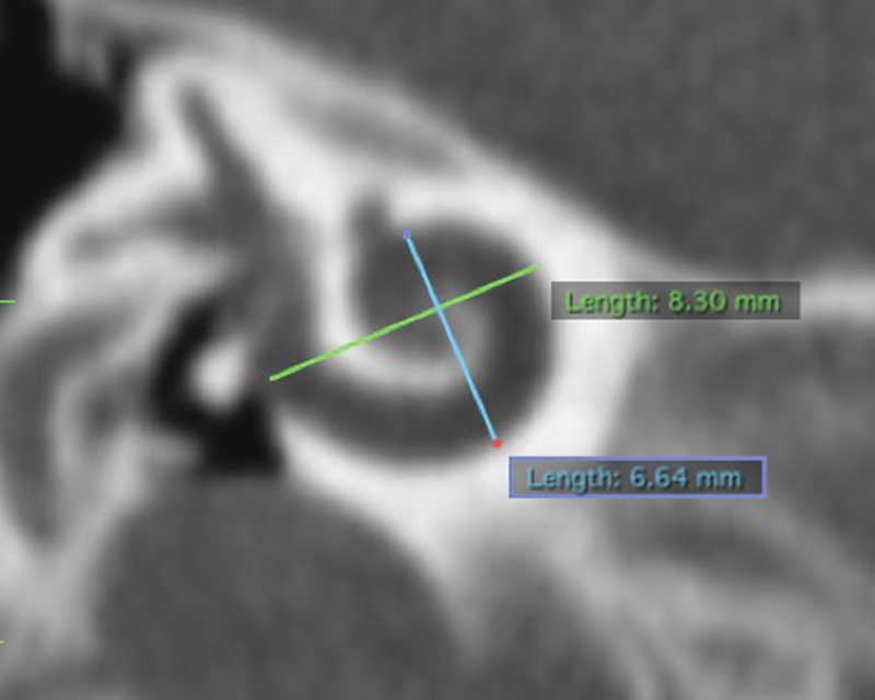
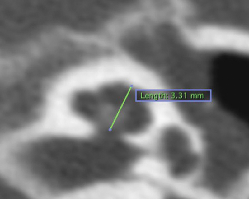
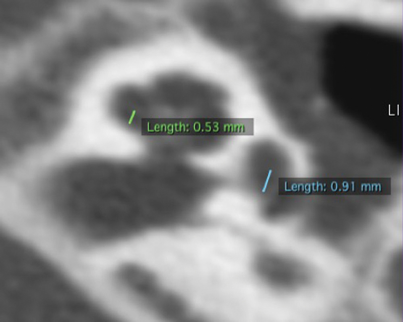
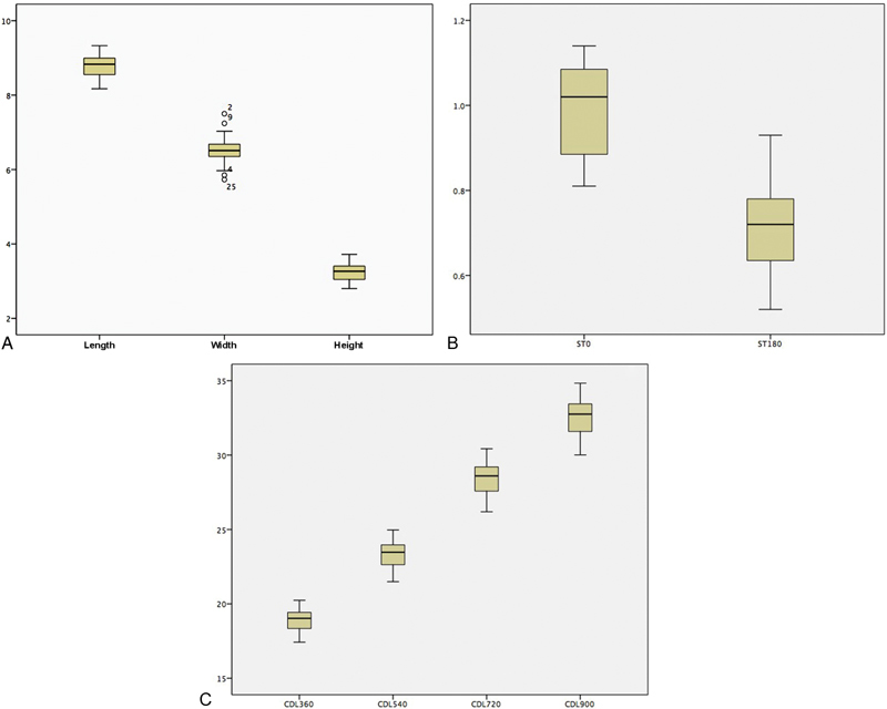
Introduction The cochlear anatomy varies in each individual, and that has an impact on decisions regarding the insertion of electrodes. The measurement of the cochlear size is the routine examination required to choose the proper cochlear implant (CI) electrodes. Objective To acquire normative data on the size of the cochlea (length, width, height, scala timpani [ST] height, cochlear duct length [CDL]) of CI candidates in Medan, Indonesia. Methods This descriptive study was conducted based on high-resolution computed tomography (HRCT) temporal bone data and on HRCT temporal data manipulated to reconstruct three-dimensional (3D) multiplanar images with OsiriX MD DICOM Viewer version 9.5.1 (Pixmeo SARL, Bernex, Geneva, Switzerland) viewer of 18 patients (36 ears) who were CI candidates in Medan, Indonesia, in order to determine cochlear length (A), cochlear width, cochlear height, ST height and CDL, calculated through a simple mathematical function. Results The average cochlear length (A) was 8.75 mm (standard deviation [SD] = 0.31 mm); the average cochlear width was 6.53 mm (SD = 0.35 mm); the average cochlear height was 3.26 mm (SD = 0.24 mm) and the average ST height at the basal cochlea was 1.00 mm (SD = 0.1 mm); and 0.71 mm (SD = 0.1 mm) at the half turn of cochlea. The average total CDL was 32.45 mm (SD = 1.31 mm; range: 30.01-34.83 mm). Conclusion The cochlear size varies in each individual; therefore, the temporal bone measurement of CI candidates using HRCT is essential: for the selection of suitable implant electrodes; to minimize cochlear damages at the insertion of the electrode arrays; and to maximize the hearing improvements.
Keywords: cochlea; cochlear implant; computed tomography; temporal bone.
Figures
References
-
- Chi D H, Sabo D L. Philadelphia: Lippincott Williams & Wilkins, a Wolters Kluwer business; 2014. Pediatric audiology and implantable hearing devices; pp. 1507–1522.
-
- Xu J, Xu S A, Cohen L T, Clark G M. Cochlear view: postoperative radiography for cochlear implantation. Am J Otol. 2000;21(01):49–56. - PubMed
-
- Dimopoulos P, Muren C. Anatomic variations of the cochlea and relations to other temporal bone structures. Acta Radiol. 1990;31(05):439–444. - PubMed
LinkOut - more resources
Full Text Sources