

doi: 10.1155/2019/8973825.
eCollection 2019.
Analysis of Enamel Loss by Prophylaxis and Etching Treatment in Human Tooth Using Optical Coherence Tomography: An In Vitro Study
Affiliations
- PMID: 30956785
- PMCID: PMC6431396
- DOI: 10.1155/2019/8973825
Item in Clipboard
Analysis of Enamel Loss by Prophylaxis and Etching Treatment in Human Tooth Using Optical Coherence Tomography: An In Vitro Study
J Healthc Eng.
.
Abstract
Bonding of braces is an essential part in contemporary orthodontic treatment. For the proper strength of bracket bonding, enamel conditioning or surface treatment on tooth surface is required. Treatment on the tooth surface such as prophylaxis smoothing with pumice and enamel etching results in considerable damages to the enamel surface of the tooth. In this study, we have proposed optical coherence tomography as a noninvasive imaging technique for the evaluation of damage induced during such treatment procedures. Using depth intensity analysis of the obtained cross-sectional images, the damage resulting to the enamel surface was studied after prophylaxis smoothening and etching steps.
Figures
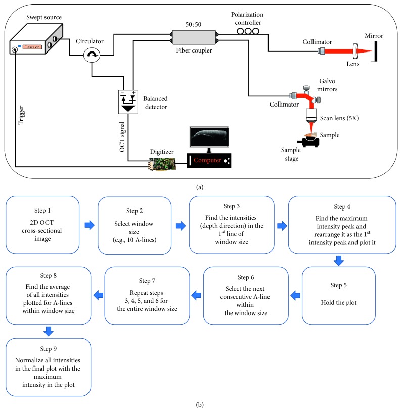
(a) The schematic diagram representing the hardware setup of the SS-OCT system. (b) The flow diagram showing the steps in depth intensity analysis algorithm.
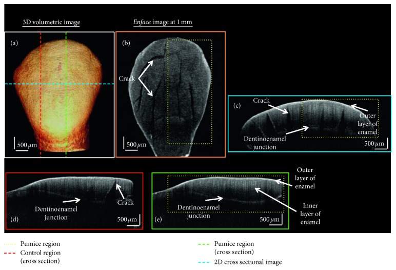
OCT images of prophylactically treated tooth sample. (a) 3D volumetric OCT image. (b) The en face image at 1 mm. (c) A 2D cross-sectional OCT image. (d) Sagittal plane showing the 2D cross-sectional image in the control region. (e) Sagittal plane showing the 2D cross-sectional image in the prophylactically treated region.
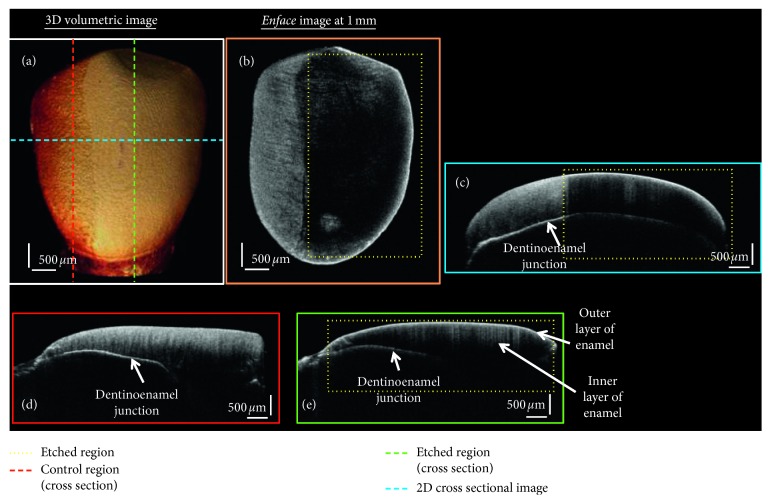
OCT images of the 37% orthophosphoric etch treated tooth sample. (a) 3D volumetric OCT image. (b) The en face image at 1 mm. (c) A 2D cross-sectional OCT image. (d) Sagittal plane showing the 2D cross-sectional image in the control region. (e) Sagittal plane showing the 2D cross-sectional image in the etch treated region.
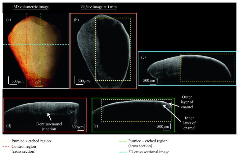
OCT images of the prophylactic and etch treated tooth sample. (a) 3D volumetric OCT image. (b) The en face image at 1 mm. (c) A 2D cross-sectional OCT image. (d) Sagittal plane showing the 2D cross-sectional image in the control region. (e) Sagittal plane showing the 2D cross-sectional image in the pumice and etch treated region.
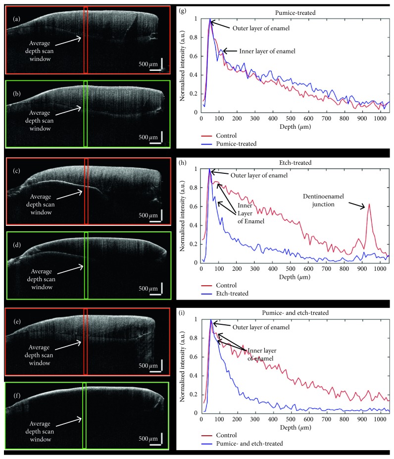
OCT cross-sectional images and the corresponding depth intensity plots of all three groups. (a, b, and g) The OCT images of prophylactically treated tooth sample and its respective depth intensity plots. (c, d, and h) The OCT images of the 37% orthophosphoric etch treated tooth sample and its respective depth intensity plots. (e, f, and i) The OCT images prophylactic and etch treated tooth sample and its respective depth intensity plots.
References
-
- Burgess A. M., Sherriff M., Ireland A. J. Self-etching primers: is prophylactic pumicing necessary? A randomized clinical trial. Angle Orthodontist. 2006;76(1):114–118. - PubMed
Publication types
MeSH terms
Substances
LinkOut - more resources
Full Text Sources
