. 2019 Mar 6:2019:3727635.
doi: 10.1155/2019/3727635. eCollection 2019.

A Rat Model of Radiation Vasculitis for the Study of Mesenchymal Stem Cell-Based Therapy

Affiliations

A Rat Model of Radiation Vasculitis for the Study of Mesenchymal Stem Cell-Based Therapy

Jian Zhang et al. Biomed Res Int. .

Abstract

Radiation vasculitis is one of the most common detrimental effects of radiotherapy for malignant tumors. This is developed at the vasculature of adjacent organs. Animal experiments have showed that transplantation of mesenchymal stem cells (MSCs) restores vascular function after irradiation. But the population of MSCs being engrafted into irradiated vessels is too low in the conventional models to make assessment of therapeutic effect difficult. This is presumably because circulating MSCs are dispersed in adjacent tissues being irradiated simultaneously. Based on the assumption, a rat model, namely, RT (radiation) plus TX (transplantation), was established to promote MSC homing by sequestering irradiated vessels. In this model, a 1.5 cm long segment of rat abdominal aorta was irradiated by 160kV X-ray at a single dose of 35Gy before being procured and grafted to the healthy counterpart. F344 inbred rats served as both donors and recipients to exclude the possibility of immune rejection. A lead shield was used to confine X-ray delivery to a 3 cm×3 cm square-shaped field covering central abdominal region. The abdominal viscera especially small bowel and colon were protected from irradiation by being pushed off the central abdominal cavity. Typical radiation-induced vasculopathy was present on the 90th day after irradiation. The recruitment of intravenously injected MSCs to irradiated aorta was significantly improved by using the RT-plus-TX model as compared to the model with irradiation only. Generally, the RT-plus-Tx model promotes MSC recruitment to irradiated aorta by separating irradiated vascular segment from adjacent tissue. Thus, the model is preferred in the study of MSC-based therapy for radiation vasculitis when the evaluation of MSC homing is demanding.


Figures

Figure 1
Figure 1
Animal groups and treatment protocols. Sixty-four female F344 rats were allocated to six groups. The RT-plus-TX and RT-plus-TX + MSC group each had eight pairs of rats, and the other groups each had eight rats. Aorta irradiation was conducted in four groups: RT-only, RT-only + MSC, RT-plus-TX, and RT-plus-TX + MSC groups. The TX-only and vehicle groups were not irradiated and served as negative control. After irradiation, the aortas from RT-plus-TX and RT-plus-TX + MSC groups were transplanted to healthy rats. The RT-only and RT-only + MSC groups served as negative control for aorta transplantation. The mesenchymal stem cells were infused to RT-only + MSC and RT-plus-TX + MSC groups starting from thirty days after irradiation for four times with the interval of fifteen days. All rats were sacrificed on the ninetieth day after irradiation, and the aortas were procured for histology and biomedical analysis.
Figure 2
Figure 2
Schematic diagram of ionizing radiation device, lead shield, and irradiated field. (a) The rats were fixed in the sterilized container after anesthesia. The abdominal cavity was opened to expose the abdominal aorta in the center of abdomen. The X-ray beam was limited to 3cm×3cm square-shaped area of the central abdomen while another part of body was protected by a customized lead shield. After irradiation, a 1.5 cm long aorta graft was procured for transplantation. (b) The aorta together with the adjacent tissues was exposed to radiation through a square-shaped orifice on the top of lead shielding container. The irradiated adjacent tissues consisted of vena cava, spine, skin, and muscle in posterior abdominal wall. (c) The thickness of rat posterior abdominal wall was estimated at 1.5cm on average.
Figure 3
Figure 3
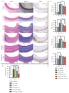
Histology and cytokine analysis of abdominal aortas. (a) Cross-sectional images of abdominal aorta. Serial cross sections of aorta were processed with hematoxylin-eosin stain (H&E), Masson's trichrome stain (Masson), and immunostaining with antimyeloperoxidase antibody using DAB substrate kit (MPO), respectively. The images represented the investigation of eight rats for each group. Scar bar 100 μm. (b) Histological analysis of intimal hyperplasia. The relative intimal thickness was normalized to full thickness of vascular wall and expressed as a percentage. Eight rats were investigated for each group. Group comparison was performed with Mann–Whitney U test. P < 0.05. (c) Expression of proinflammatory cytokines in aortas. The cytokine levels were measured by real-time qualitative reverse transcription PCR and normalized to vehicle control. The experiment was repeated three times for each group. Group comparison was performed with Mann–Whitney U test. P < 0.05.
Figure 4
Figure 4
Engraftment of mesenchymal stem cells (MSC) in irradiated aorta. MSC engraftment was investigated by two techniques: fluorescent microscopy for tracing cells with green fluorescent protein (GFP) label and PCR analysis for sex determination region on the Y chromosome (Sry) specifically carried by transplanted MSCs. For each group, eight rats were investigated by fluorescent microscopy, and the PCR analysis of aortic tissue homogenates from eight rats was repeated three times. Group comparison was performed with Mann–Whitney U test. P < 0.05.
Figure 5
Figure 5
Illustrative mechanism by which RT-plus-TX model promotes the engraftment of mesenchymal stem cells (MSCs). After local irradiation, the aortas together with adjacent tissues are subjected to injury and release damage signal to attract the migration of infused MSCs. The migrated MSCs are dispersed in the aortas and adjacent tissues, leading to seemly low frequency of MSC engraftment. When the irradiated aortas are transplanted to isogenic healthy rats, the injured aortas live with normal surrounding tissues. The MSCs are prone to gather in the injured aortas, and the frequency of MSC engraftment is improved.

Similar articles

Cited by

References

    1. Chen J., Yen Y., Liu S., et al. Dementia risk in irradiated patients with head and neck cancer. Medicine. 2015;94(45):p. e1983. doi: 10.1097/MD.0000000000001983. - DOI - PMC - PubMed
    1. Chong V. F. H., Khoo J. B. K., Chan L. L., Rumpel H. Neurological changes following radiation therapy for head and neck tumours. European Journal of Radiology. 2002;44(2):120–129. doi: 10.1016/s0720-048x(02)00066-9. - DOI - PubMed
    1. Xu J., Cao Y. Radiation-induced carotid artery stenosis: a comprehensive review of the literature. Interventional Neurology. 2013;2(4):183–192. doi: 10.1159/000363068. - DOI - PMC - PubMed
    1. Harb A. H., Abou Fadel C., Sharara A. I. Radiation enteritis. Current Fungal Infection Reports. 2014;16(5):p. 383. - PubMed
    1. Fajardo L. F. The pathology of ionizing radiation as defined by morphologic patterns. Acta Oncologica. 2005;44(1):13–22. doi: 10.1080/02841860510007440. - DOI - PubMed