. 2019 Apr 30;58(17):2269-2281.
doi: 10.1021/acs.biochem.9b00123. Epub 2019 Apr 18.

Ca(II) and Zn(II) Cooperate To Modulate the Structure and Self-Assembly of S100A12

Affiliations

Ca(II) and Zn(II) Cooperate To Modulate the Structure and Self-Assembly of S100A12

Qian Wang et al. Biochemistry. .

Abstract

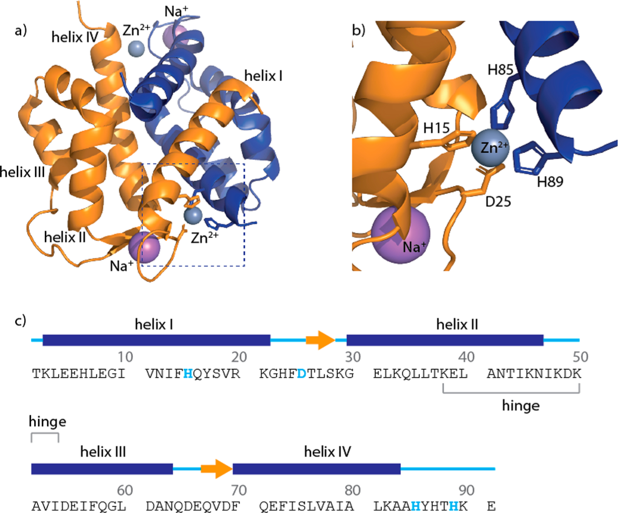
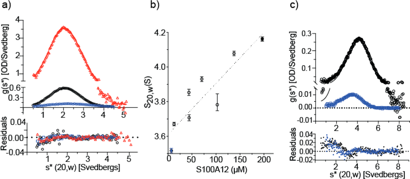
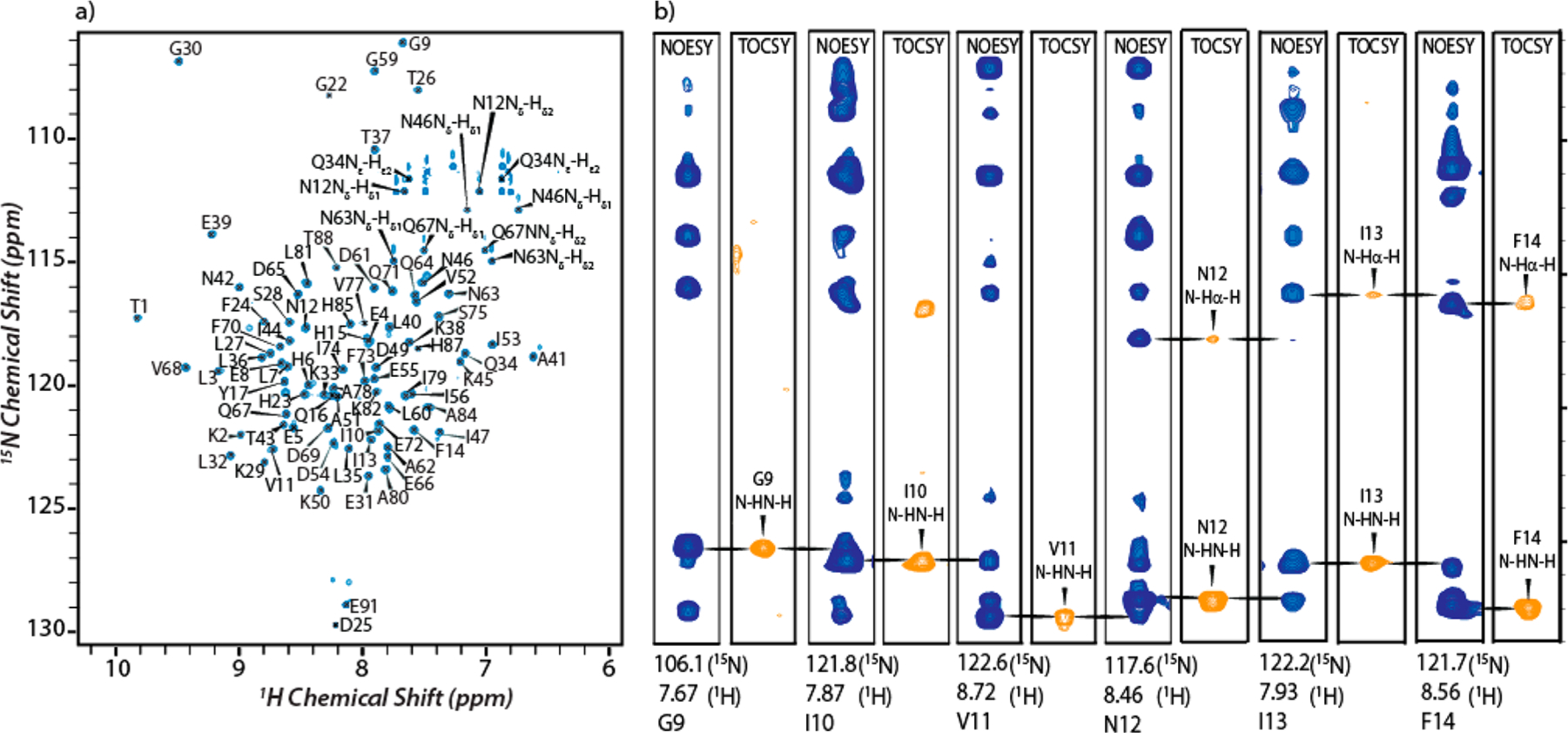
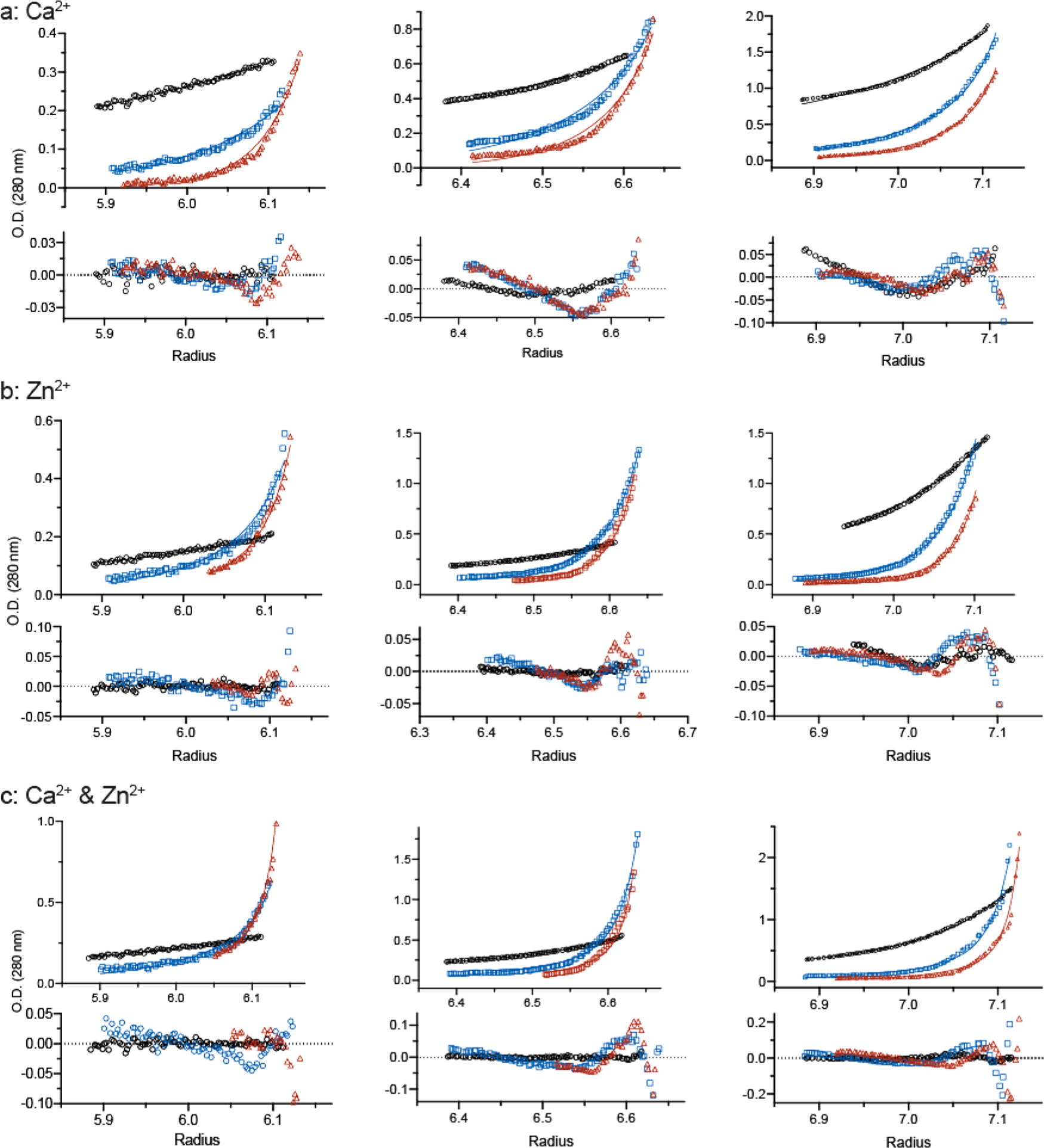
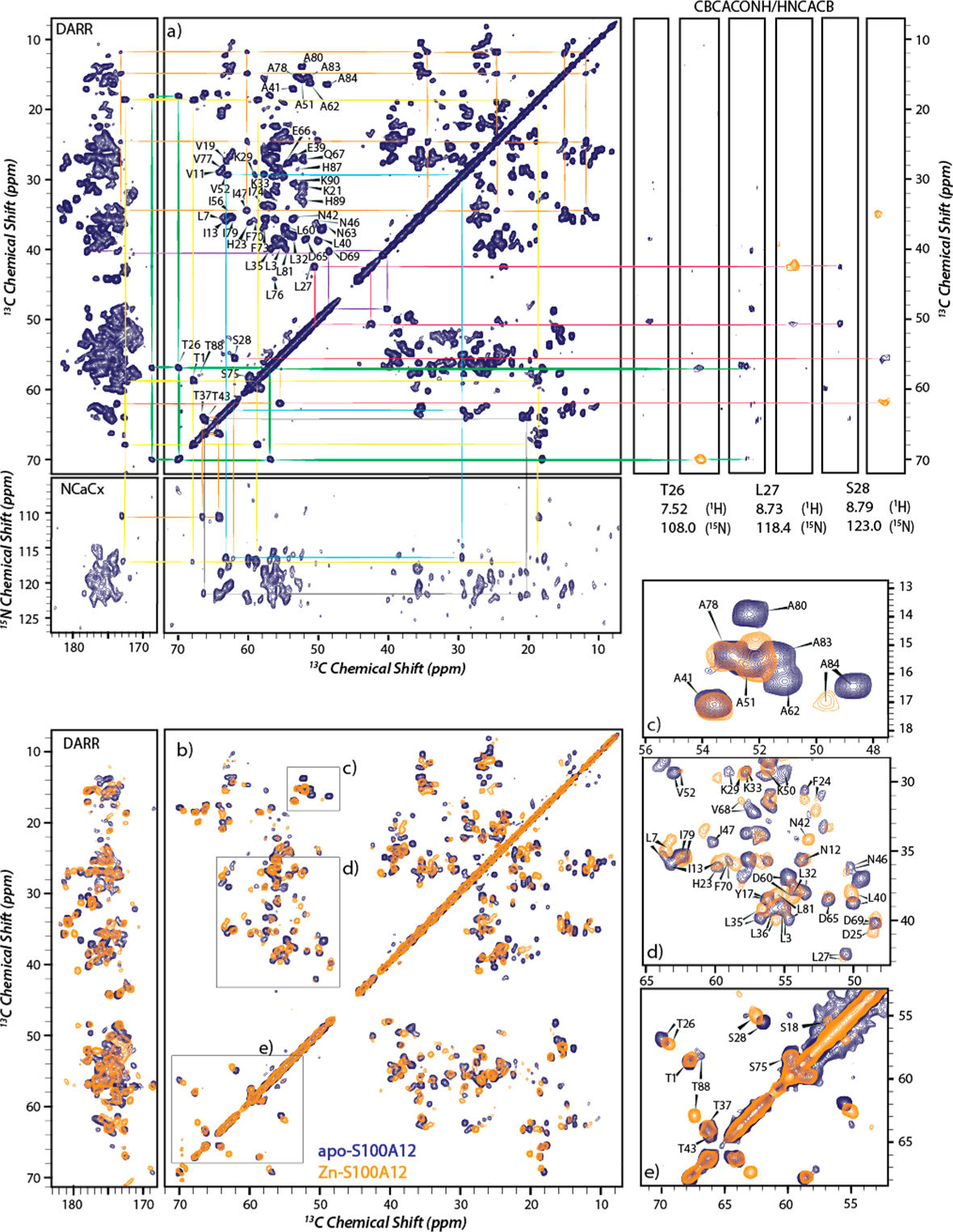
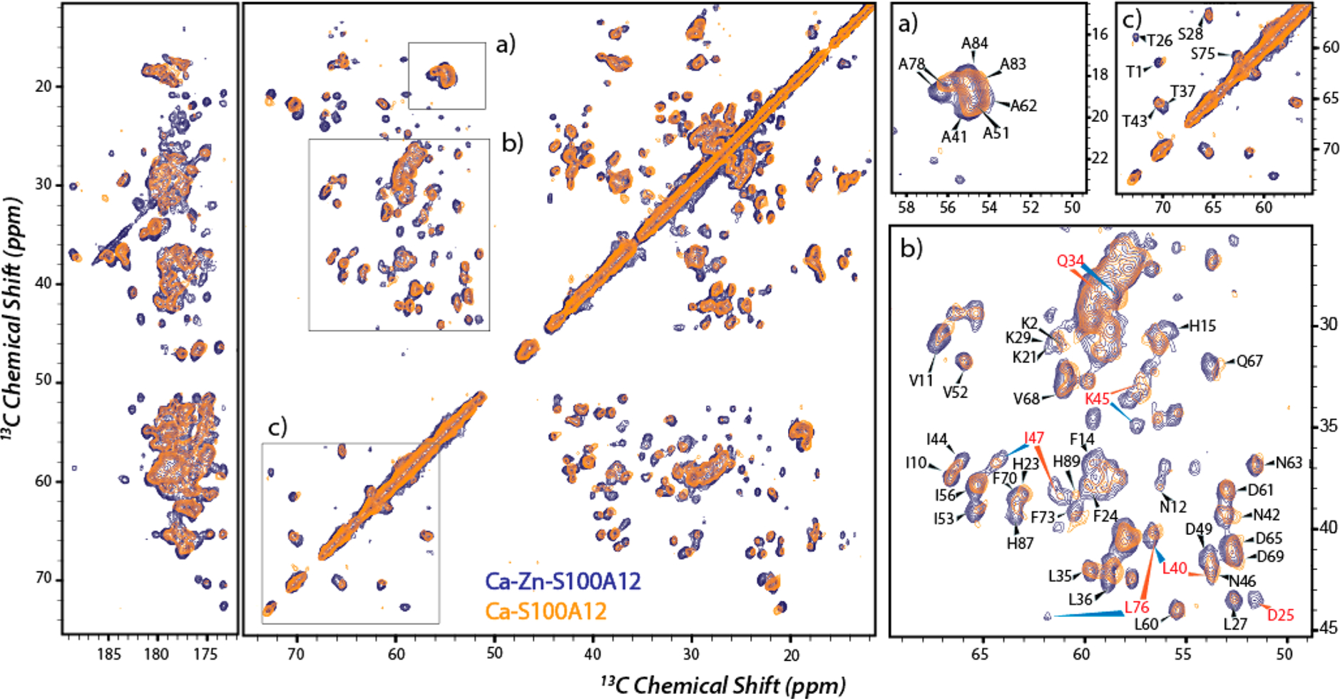
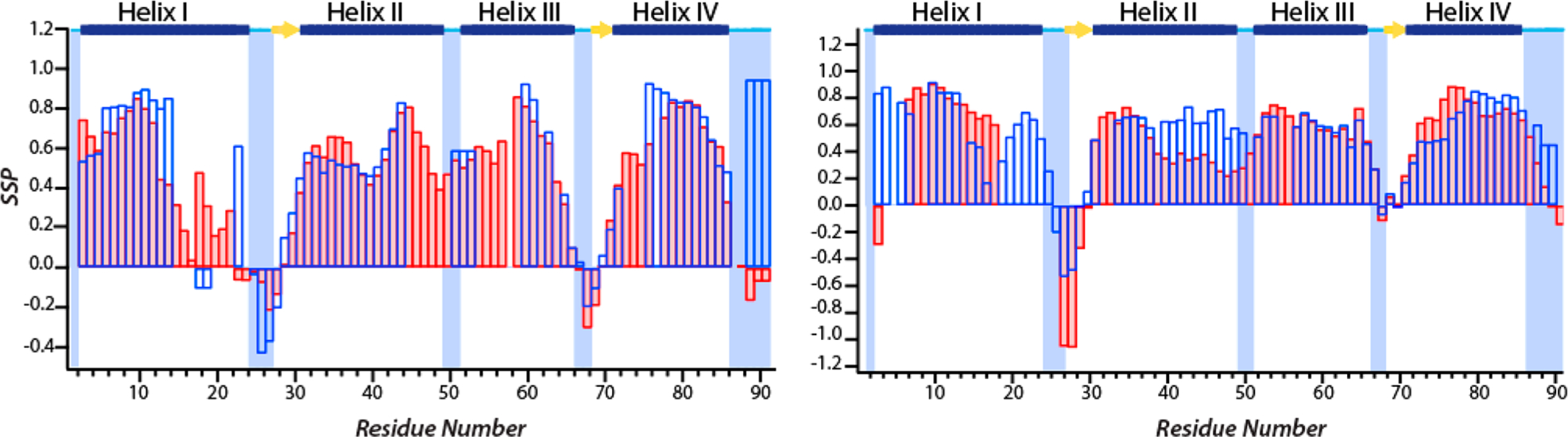
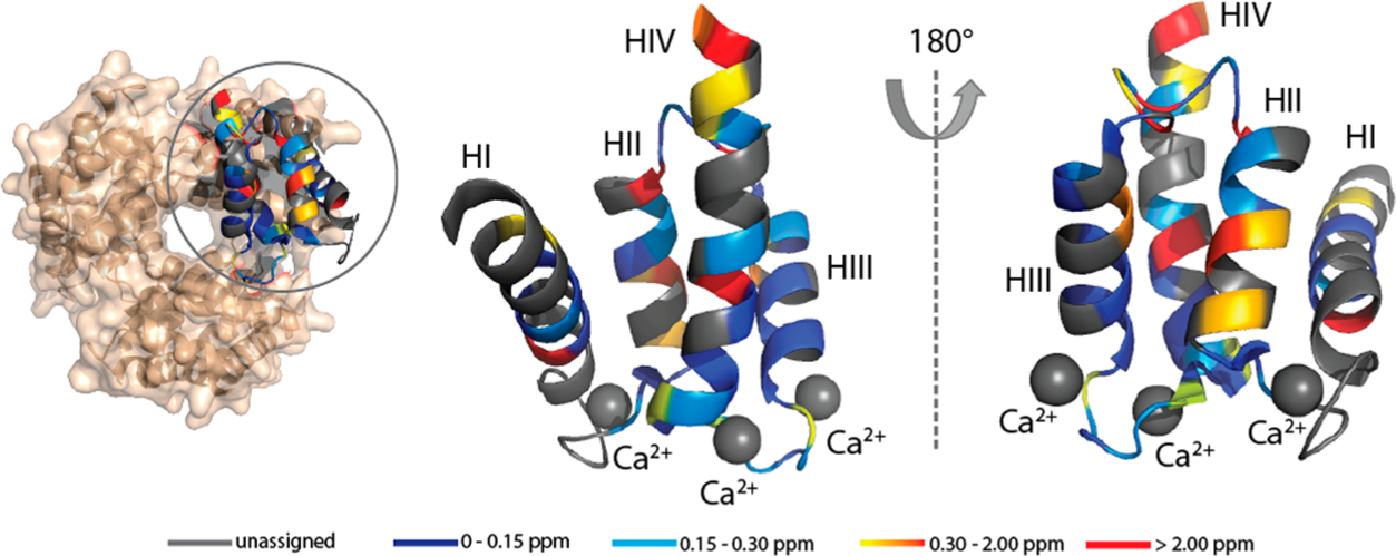
S100A12 is a member of the Ca2+ binding S100 family of proteins that functions within the human innate immune system. Zinc sequestration by S100A12 confers antimicrobial activity when the protein is secreted by neutrophils. Here, we demonstrate that Ca2+ binding to S100A12's EF-hand motifs and Zn2+ binding to its dimeric interface cooperate to induce reversible self-assembly of the protein. Solution and magic angle spinning nuclear magnetic resonance spectroscopy on apo-, Ca2+-, Zn2+-, and Ca2+,Zn2+-S100A12 shows that significant metal binding-induced chemical shift perturbations, indicative of conformational changes, occur throughout the polypeptide chain. These perturbations do not originate from changes in the secondary structure of the protein, which remains largely preserved. While the overall structure of S100A12 is dominated by Ca2+ binding, Zn2+ binding to Ca2+-S100A12 introduces additional structural changes to helix II and the hinge domain (residues 38-53). The hinge domain of S100A12 is involved in the molecular interactions that promote chemotaxis for human monocyte, acute inflammatory responses and generates edema. In Ca2+-S100A12, helix II and the hinge domain participate in binding with the C-type immunoglobulin domain of the receptor for advanced glycation products (RAGE). We discuss how the additional conformational changes introduced to these domains upon Zn2+ binding may also impact the interaction of S100A12 and target proteins such as RAGE.


Conflict of interest statement

The authors declare no competing financial interest.

Figures

Figure 1.
Figure 1.
Amino acid sequence and structure of zinc-bound S100A12. (a) Crystal structure of Zn2+-S100A12 (PDB entry 2WCB). (b) Close-up showing the zinc binding motif with residues H15 and D25 (orange) from one monomer and H85 and H89 (blue) from the second monomer. (c) Polypeptide sequence and secondary structure of the apoprotein based on the crystal structure (PDB entry 2WCF). Gray and purple spheres denote zinc and sodium ions, respectively.
Figure 2.
Figure 2.
Sedimentation velocity (SV) analyses of (a) apo-S100A12 and (b and c) Zn2+-S100A12 with the cation present in 2-fold molar excess relative to the protein. Panels A and C show the time-derivative distributions and the best fits of the data to a single-component (normal) distribution (Table 1 for panel A). In panel A, apo-S100A12 was analyzed at 233 μM (red), 23 μM (black), and 6 μM (blue). Sedimentation of the first sample was tracked at 280 nm; the latter samples were tracked at 230 nm. Panel C shows two of the Zn2+-S100A12 samples that were analyzed: 196 μM (black) and 6.9 μM (blue) tracked at 280 and 230 nm, respectively. Panel B shows that S20,w increases with S100A12 concentration for the eight protein concentrations analyzed; the solid blue and black symbols represent the two distributions shown in panel C. The dotted line depicts a linear regression that highlights the upward trend of the data and does not represent a particular assembly model.
Figure 3.
Figure 3.
1H–15N HSQC spectrum and selected two-dimensional planes of the 3D 1H–15N NOESY-HSQC and TOCSY-HSQC spectra: (a) 14.1 T 1H–15N HSQC spectrum of apo-S100A12 with assignment of resonances and (b) strip plots from 1H–15N NOESY-HSQC (blue) and TOCSYHSQC (orange) spectra for residues G9–F14.
Figure 4.
Figure 4.
Sedimentation equilibrium analysis of S100A12 equilibrated in a buffer containing (a) 20 mM Ca2+, (b) 0.2 mM Zn2+, and (c) 20 mM Ca2+ and 0.2 mM Zn2+. The protein concentrations loaded into each sector of the six-channel centerpiece were 33 μM (left), 98 μM (middle), and 327 μM (right). The concentration gradients were determined using the absorption optics set at 280 nm. The best global fits of the dimer–octamer model to the three protein concentrations equilibrated at 12000 (black), 24000 (blue), and 30000 rpm (red) are shown along with the residuals; the resolved Kd values are cited in the text.
Figure 5.
Figure 5.
MAS NMR spectra of apo- and Zn2+-S100A12. (a) 13C–13C DARR (14.1 T) and 13C–15N NCACX (16.4 T) 2D correlation spectra of apo-S100A12 with resonance assignments (left) and strip plots from 1H–13C–15N CBCACONH (blue) and HNCACB (orange) solution NMR spectra for residues T26–S28 (right). Signals belonging to an assigned residue in MAS and solution spectra are connected with the same color. (b) Overlay of Zn2+-S100A12 (orange) and apo-S100A12 (blue) 13C–13C DARR MAS spectra acquired at 14.1 T. (c–e) Close-ups of panel b with assignments of the perturbed residues. All solid-state NMR spectra were acquired at 12 kHz MAS.
Figure 6.
Figure 6.
Overlay of 13C–13C correlation MAS NMR spectra of Ca2+,Zn2+-S100A12 (blue) and Ca2+-S100A12 (orange). Panels a–c are close-ups with assignments showing perturbed residues. Residues marked in red show significant CSP upon zinc binding. The experimental conditions were the same as those described for Figure 5.
Figure 7.
Figure 7.
Site-specific CSPs for binding of zinc to apo-S100A12 (left) and Ca2+-S100A12 (right). Cα, NH, and Cβ perturbations are colored pink, yellow, and blue, respectively. The secondary structure of the apoprotein determined from the crystal structure (PDB entry 2WCF) is displayed at the top: α-helices (blue bars), β-strands (yellow arrows), and loop regions (cyan lines). The dashed horizontal lines represent CSPs of 0.15 and 0.3 ppm for Cα and Cβ and 0.50 and 1.00 ppm for NH.
Figure 8.
Figure 8.
Comparison of the SSPs predicted from Cα and Cβ chemical shifts: apo-S100A12 (pink bars) and Zn2+-S100A12 (empty blue bars) (left) and Ca2+-S100A12 (pink bars) and Ca2+,Zn2+-S100A12 (empty blue bars) (right). The secondary structure of the apoprotein and Ca2+-bound protein determined from the crystal structure (PDB entries 2WCF and 1E8A) is displayed at the top: α-helices (blue bars), β-strands (yellow arrows), and loop regions (cyan lines).
Figure 9.
Figure 9.
Observed Cα CSPs mapped onto the crystal structure of apo-S100A12 (PDB entry 2WCF). Color scheme: gray for unassigned residues, blue for residues with CSPs between 0 and 0.15 ppm, cyan for residues with CSPs between 0.15 and 0.3 ppm, gradient from orange to red for residues with CSPs between 0.30 and 2.00 ppm, and red for residues with CSPs of >2.00 ppm. The zinc binding residues are shown as sticks and labeled in red.
Figure 10.
Figure 10.
Observed Cα CSPs observed upon binding of Zn2+ to Ca2+-S100A12 mapped onto the crystal structure of hexameric S100A12 (PDB entry 1GQM). The CSPs are mapped on the monomeric chain for the sake of clarity. Color scheme: gray for unassigned residues, blue for residues with CSPs between 0 and 0.15 ppm, cyan for residues with CSPs between 0.15 and 0.3 ppm, gradient from orange to red for residues with CSPs between 0.30 and 2.00 ppm, and red for residues with CSPs of >2.00 ppm. The calcium ions are denoted by gray spheres.

References

    1. Hood MI, and Skaar EP (2012) Nutritional immunity: transition metals at the pathogen–host interface. Nat. Rev. Microbiol 10, 525–537. - PMC - PubMed
    1. Kehl-Fie TE, and Skaar EP (2010) Nutritional immunity beyond iron: a role for manganese and zinc. Curr. Opin. Chem. Biol 14, 218–224. - PMC - PubMed
    1. Weinberg ED (1975) Nutritional immunity: host’s attempt to withhold iron from microbial invaders. JAMA, J. Am. Med. Assoc 231, 39–41. - PubMed
    1. Zackular JP, Chazin WJ, and Skaar EP (2015) Nutritional immunity: S100 proteins at the host-pathogen interface. J. Biol. Chem 290, 18991–8. - PMC - PubMed
    1. Nakashige TG, Zygiel EM, Drennan CL, and Nolan EM (2017) Nickel sequestration by the host-defense protein human calprotectin. J. Am. Chem. Soc 139, 8828–8836. - PMC - PubMed

Publication types