. 2019 Feb 28;16(151):20180678.
doi: 10.1098/rsif.2018.0678.

Knee implant kinematics are task-dependent

Affiliations

Knee implant kinematics are task-dependent

Pascal Schütz et al. J R Soc Interface. .

Abstract

Although total knee arthroplasty (TKA) has become a standard surgical procedure for relieving pain, knowledge of the in vivo knee joint kinematics throughout common functional activities of daily living is still missing. The goal of this study was to analyse knee joint motion throughout complete cycles of daily activities in TKA subjects to establish whether a significant difference in joint kinematics occurs between different activities. Using dynamic videofluoroscopy, we assessed tibio-femoral kinematics in six subjects throughout complete cycles of walking, stair descent, sit-to-stand and stand-to-sit. The mean range of condylar anterior-posterior translation exhibited clear task dependency across all subjects. A significantly larger anterior-posterior translation was observed during stair descent compared to level walking and stand-to-sit. Local minima were observed at approximately 30° flexion for different tasks, which were more prominent during loaded task phases. This characteristic is likely to correspond to the specific design of the implant. From the data presented in this study, it is clear that the flexion angle alone cannot fully explain tibio-femoral implant kinematics. As a result, it seems that the assessment of complete cycles of the most frequent functional activities is imperative when evaluating the behaviour of a TKA design in vivo.

Keywords: activities of daily living; gait activities; moving fluoroscope; tibio-femoral kinematics; total knee arthroplasty; videofluoroscopy.


Conflict of interest statement

R.L. has received speaker's fees from Medacta and DePuy Synthes. R.L. has received research funding from DePuy Synthes and Medacta International.

Figures
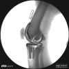
Figure 1.
Figure 1.
Exemplary instant of three-dimensional tibial and femoral components registered to the two-dimensional fluoroscopic image. The two-dimensional/three-dimensional registration of all images allows the internal joint kinematics to be determined over the complete cycles of functional activities.
Figure 2.
Figure 2.
Implant coordinate systems for the femoral and tibial components (a) and nearest points for stance (red) and swing (green) phase for an exemplary trial of stair descent presented in the coordinate system of the tibial component (b).
Figure 3.
Figure 3.
Tibio-femoral flexion is presented for all tasks, including mean and SD over all subjects. Loaded and unloaded activity phases are shown, respectively, for level walking (red/orange), stair descent (green/light green), sit-to-stand (dark blue) and stand-to-sit (light blue).
Figure 4.
Figure 4.
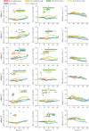
Tibio-femoral kinematics are shown for the activities of level walking, stair descent, sit-to-stand and stand-to-sit for each single subject, represented by the different colours, including the mean and SD over all repetitions. The columns are presented in % cycle and show the A–P translation of the medial and lateral condyles, as well as the tibial internal/external rotation, and joint ab/adduction. For level walking and stair descent, the dashed lines indicate the mean time point of toe-off for each subject.
Figure 5.
Figure 5.
A–P translation for the medial (a) and lateral (b) condyles, as well as tibial rotation (c) are shown as a function of the flexion angle. The tasks level walking, stair descent, sit-to-stand and stand-to-sit are presented as the mean of all six subjects, with the group SD shown as transparent. Note that an anterior translation of the nearest point on the medial condyle and/or posterior translation on the lateral condyle represents an internal rotation of the tibial component. Asterisk (*): significant differences, found between the tasks for certain ranges of flexion, were indicated with bars in the colours of the respective tasks with an adjusted level of significance of α = 0.0033.
Figure 6.
Figure 6.
Average positions across all repetitions of the femoral component, represented by lines connecting the nearest points of the medial and lateral condyles relative to the tibial tray for specific time points (dark to bright) during the cycles of level walking (red tones), stair descent (green tones), sit-to-stand (dark blue tones) and stand-to-sit (bright blue tones). Solid lines represent the loaded stance phase and dotted lines the unloaded swing phase.
Figure 7.
Figure 7.
A–P translation for the medial and lateral condyles, as well as tibial rotation are shown as a function of the flexion angle. The tasks level walking, stair descent, sit-to-stand and stand-to-sit are presented for each of the six subjects as the mean of all repetitions, with the subject SD shown as transparent. Asterisk (*): significant differences, found between the tasks for certain ranges of flexion, were indicated with bars in the colours of the respective tasks with an adjusted level of significance of α = 0.0033.

References

    1. Robertsson O, Dunbar M, Pehrsson T, Knutson K, Lidgren L. 2000. Patient satisfaction after knee arthroplasty: a report on 27,372 knees operated on between 1981 and 1995 in Sweden. Acta Orthop. Scand. 71, 262–267. (10.1080/000164700317411852) - DOI - PubMed
    1. Fenner VU, Behrend H, Kuster MS. 2017. Joint mechanics after total knee arthroplasty while descending stairs. J. Arthroplasty 32, 575–580. (10.1016/j.arth.2016.07.035) - DOI - PubMed
    1. Andriacchi TP, Galante JO, Fermier RW. 1982. The influence of total knee-replacement design on walking and stair-climbing. J. Bone Joint Surg. Am. 64, 1328–1335. (10.2106/00004623-198264090-00008) - DOI - PubMed
    1. Trinler UK, Baty F, Mundermann A, Fenner V, Behrend H, Jost B, Wegener R. 2016. Stair dimension affects knee kinematics and kinetics in patients with good outcome after TKA similarly as in healthy subjects. J. Orthop. Res. 34, 1753–1761. (10.1002/jor.23181) - DOI - PubMed
    1. Taylor WR, Ehrig RM, Duda GN, Schell H, Seebeck P, Heller MO. 2005. On the influence of soft tissue coverage in the determination of bone kinematics using skin markers. J. Orthop. Res. 23, 726–734. (10.1016/j.orthres.2005.02.006) - DOI - PubMed

Publication types