. 2019 Apr 20;132(8):948-956.
doi: 10.1097/CM9.0000000000000162.

Impact of Taurine on the proliferation and apoptosis of human cervical carcinoma cells and its mechanism

Affiliations

Impact of Taurine on the proliferation and apoptosis of human cervical carcinoma cells and its mechanism

Hua Li et al. Chin Med J (Engl). .

Abstract

Background: Cervical cancer has the fourth highest incidence and mortality rate of all cancers in women worldwide; it seriously harms their physical and mental health. The aim of this study was to observe the roles and preliminary mechanism of Taurine (Tau)-induced apoptosis in cervical cancer cells.

Methods: Cells from the human cervical cancer cell line SiHa were transfected with the recombinant plasmid pEGFP-N1-MST1 (mammalian sterile 20-like kinase 1); then, the cell proliferation activity was analyzed by the MTT assay, cell apoptosis by flow cytometry, and the related protein levels by Western blotting.

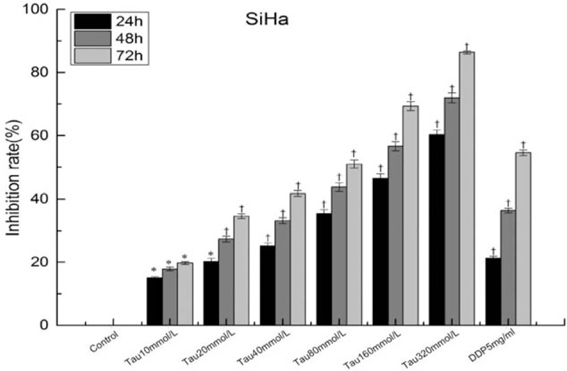
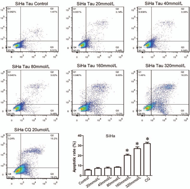
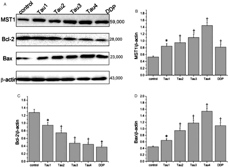
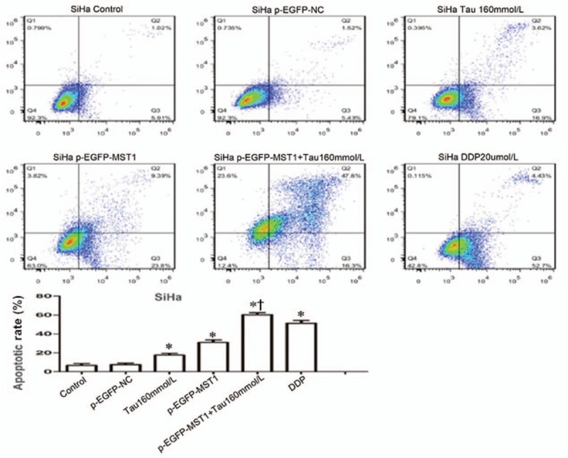
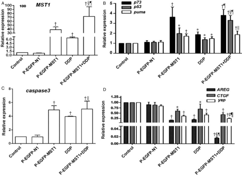
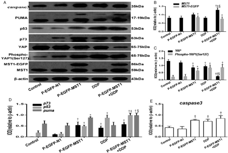
Results: Tau inhibited the proliferation of SiHa cells and induced apoptosis in these cells (the apoptotic rate was 21.95% in the Tau 160 mmol/L group and 30% in the Tau 320 mmol/L group), upregulated the expression of the MST1 (control, 0.53; Tau 40-320 mmol/L groups, 0.84-1.45) and Bax (control, 0.45; Tau 40-320 mmol/L groups, 0.64-1.51) proteins (P < 0.01), and downregulated the expression of Bcl-2 (control, 1.28, Tau 40-320 mmol/L groups, 0.93-0.47) (P < 0.01). The overexpression of MST1 promoted the apoptosis of SiHa cells, enhanced the apoptosis-inductive effects of Tau (P < 0.01), upregulated the expression of the proapoptotic proteins p73, p53, PUMA (p53 upregulated modulator of apoptosis), and caspase-3, and promoted the phosphorylation of YAP (Yes-associated protein).

Conclusions: Tau inhibited the proliferation and induced the apoptosis of cervical cancer SiHa cells. The MST1 protein plays an important role in the Tau-induced apoptosis of cervical cancer cells.


Figures

Figure 1
Figure 1
Inhibition of Tau on the proliferation of cervical cancer cells. P < 0.05 vs. Control group, P < 0.01 vs. Control group.
Figure 2
Figure 2
Effects of Tau on the apoptosis of cervical cancer cells. The SiHa cells were treated by Tau with different concentrations 0 (control), 20, 40, 80, 160, 320 mmol/L Tau, and 20 μmmol/L DDP for 48 h, and the apoptosis of SiHa cells was detected by the Annexin-V-FITC/PI double staining method. DDP is the positive control. P < 0.01 vs. the Control group.
Figure 3
Figure 3
Effects of taurine on the expressions of MST1, Bax, and Bcl-2 protein in cervical cancer cells. (A) A representative western blotting is shown. (B-D) Relative protein expression levels of Mst1, Bax, and Bcl-2 were assessed calculating the integral optical density (IOD)-values. IOD values were normalized to those of beta-actin protein. P < 0.05 vs. Control, P < 0.01 vs. Control.
Figure 4
Figure 4
Effects of MST1 overexpression on Tau-induced apoptosis in SiHa cells. The apoptosis of SiHa cells was detected by Annexin-V-FITC/PI double staining; compared with the Control group or the p-EGFP-NC group, the apoptotic rates in group Tau 160 mmol/L, group p-EGFP-MST1, and group p-EGFP-MST1+ Tau 160 mmol/L were significantly increased (20.52%, 33.19%, and 64.1%, respectively, P < 0.01). Compared with the p-EGFP-MST1 group, the apoptotic rate in group p-EGFP-MST1+ Tau 160 mmol/L still exhibited significant increase (P < 0.01), suggesting that MST1 and Tau may have synergistic effects. P < 0.01 vs. Control or p-EGFP-NC; P < 0.01 vs. p-EGFP-MST1.
Figure 5
Figure 5
Effects of MST1 overexpression on the mRNA expressions of p73, p53, and PUMA in SiHa cells of different groups. The RT-qPCR results showed that compared with the Control group and the p-EGFP-N1 group, the expression of MST1 mRNA in the p-EGFP-MST1 group and the p-EGFP-MST1+DDP group were significantly increased by 40 times and 70 times, respectively, than the control group (A); the expressions of mRNA of p73 (P < 0.01), p53 (P < 0.05), puma (P < 0.01), and caspase3 (P < 0.01) also increased to varying degrees (B,C); the expressions of CTGF, AREG, and yap in the p-EGFP-MST1 group decreased to varying degrees; compared with the p-EGFP-MST1 group, the expressions of CTGF, AREG, and yap mRNA in the p-EGFP- MST1 + DDP group were significantly decreased (D). P < 0.05, P < 0.01 vs. Control or p-EGFP-N1; P < 0.05, §P < 0.01 vs. p-EGFP-MST1; ||P < 0.05, P < 0.01 vs. DDP.
Figure 6
Figure 6
Effects of Mst1 overexpression on the expressions of YAP, p73, p53, PUMA, and caspase-3 protein. The results of Western blot show that compared with the Control group or the p-EGFP-N1 group, the total MST1 protein expression in the cells was increased by 2.2 times after transfection of p-EGFP-MST1 plasmid (A and B); The YAP protein was significantly decreased (P < 0.01); the Phospho-YAP protein was significantly increased, and the ratio of Phospho-YAP1(Ser127) to YAP was increased by about 4.5 times (A and C); the expressions of p73, p53, PUMA and caspase -3 protein were significantly increased (P < 0.05 or P < 0.01) (A, D and E). Compared with the p-EGFP-MST1 group, the expressions of proapoptotic protein p73, p53, PUMA, and caspase-3 in the p-EGFP-MST1+DDP group were significantly increased (P < 0.01) (A, D and E). P < 0.05, P < 0.01 vs. Control or p-EGFP-N1; P < 0.01 vs. p-EGFP-MST1; §P < 0.01 vs. 5-DDP.

References

    1. Torre LA, Bray F, Siegel RL, Ferlay J, Lortet-Tieulent J, Jemal A. Global cancer statistics, 2012. CA Cancer J Clin 2015; 65:87–108. doi: 10.3322/caac.21262. - PubMed
    1. Chai RC, Lambie D, Verma M, Punyadeera C. Current trends in the etiology and diagnosis of HPV-related head and neck cancers. Cancer Med 2015; 4:596–607. doi: 10.1002/cam4.424. - PMC - PubMed
    1. De Sanjosé S, Bruni L, Alemany L. HPV in genital cancers (at the exception of cervical cancer) and anal cancers. Presse Med 2014; 43:e423–e428. doi: 10.1016/j.lpm.2014.10.001. - PubMed
    1. Nelson EJ, Hughes J, Kulasingam SL. Spatial patterns of human papillomavirus-associated cancers within the state of Minnesota, 1998-2007. Spat Spatiotemporal Epidemiol 2014; 9:13–21. doi: 10.1016/j.sste.2014.02.003. - PubMed
    1. Munger K. Are selective estrogen receptor modulators (SERMs) a therapeutic option for HPV-associated cervical lesions and cancers? Am J Pathol 2014; 184:358–361. doi: 10.1016/j.ajpath.2013.11.005. - PubMed

MeSH terms