. 2019 Jun;79(9):949-960.
doi: 10.1002/pros.23802. Epub 2019 Apr 8.

Optimizing live-animal bioluminescence imaging prediction of tumor burden in human prostate cancer xenograft models in SCID-NSG mice

Affiliations

Optimizing live-animal bioluminescence imaging prediction of tumor burden in human prostate cancer xenograft models in SCID-NSG mice

Sangyub Kim et al. Prostate. 2019 Jun.

Abstract

Background: Noninvasive live-animal longitudinal monitoring of xenograft tumor growth and metastasis by bioluminescent imaging (BLI) has been widely reported in cancer biology and preclinical therapy literature, mainly in athymic nude mice. Our own experience at calibrating BLI readout with tumor weight/volume in human prostate cancer xenograft models in haired, SCID-NSG mice through intraprostatic (orthotopic) and subcutaneous (SC) inoculations revealed either nonexistent or poor correlation (coefficient of determination, R 2 = ~0.01-0.3). The present work examined several technical and biological factors to improve BLI utility.

Methods: After ruling out promoter-luciferase (luc) specificity and luc gene loss in the cell inoculum with LNCaP-AR-luc cells expressing an androgen receptor (AR) and tagged with AR-responsive probasin promoter-luc gene, we evaluated different routes of d-luciferin administration, imaging time during the day, charge-coupled device camera image acquisition settings, and hair removal methods to improve the imaging protocol. For most imaging sessions, BLI was carried out within the same day of tumor volume measurement. After necropsy, histological and immunohistochemical (IHC) analyses were performed on the tumors to evaluate necrosis and expression of luciferase and AR, respectively.

Results: Injection of d-luciferin by SC route, robust image-capture setting (30 000 counts and autoexposure), imaging in the morning and thorough hair removal resulted in a substantial improvement of R2 to ~0.6. Histological analyses confirmed the lack of BLI signal in necrotic tumor masses consistent with luciferase-mediated light emission only in oxygenated adenosine triphosphate-producing viable cells. IHC staining detected heterogeneous expression of luciferase tracking generally with AR expression in nonnecrotic tumor tissues.

Conclusions: Our body of work highlighted a framework to validate imaging protocols to ensure the acquisition of interpretable BLI data as an indicator of xenograft tumor burden. The vast tissue heterogeneity in prostate tumor xenografts and variable luciferase expression constrained this technology from achieving a high correlation.

Keywords: IVIS instrumental setting; bioluminescence; heterogeneous expression of luciferase; necrosis; prostate cancer.


Conflict of interest statement

Conflict of Interest: The authors declare that they have no conflict of interest.

Figures

Fig 1.
Fig 1.
(A) Example of weekly BLI of a mouse for intra-animal comparison of BLI readouts between tumors inoculated subcutaneously on the left and right flank (TX-013 study). IP luciferin injection. (B) Correlation plot between BLI flux outputs and final tumor weights. (C) Plot of BLI flux normalized by tumor weight to compare left and right tumors in mice bearing dual tumors. (D) BLI images of a mouse (PA-04 study) measured in the morning (upper panel) and in the afternoon with IP injection failure (bottom left panel) and the repeat injection for BLI detection correction (bottom right panel).
Fig 2.
Fig 2.
Flow cytometric analyses for luciferase expression in LNCaP-AR-luc cells. (A) Negative control (no antibody). (B) Luciferase antibody alone. (C) Secondary antibody alone. (D) Luciferase antibody plus 2nd antibody. Note log scale for x-axis. All cells stained positive for luciferase protein with a range of an order of magnitude apart from low to high expressors.
Fig 3.
Fig 3.
Comparison of BLI by IV luciferin delivery in the morning vs. BLI in the afternoon with IP luciferin delivery for correlation with tumor volume (Instrument setting: 3,000 counts and 10 sec. exposure). For IV luciferin delivery, (A) BLI kinetics and correlations for (B) first and (C) second measurements were plotted. For IP luciferin delivery, (D) BLI kinetics and correlations for (E) first and (F) second measurements were plotted. Note the first and second measurements were done in separate sessions 6 days apart.
Fig 4.
Fig 4.
Comparison of correlation and intra-day reproducibility for BLI after IP or SC luciferin administration (Instrument setting: 30,000 counts and auto-exposure). For IP luciferin delivery, correlations for (A) morning and (B) afternoon measurements were plotted. For SC luciferin delivery, correlations for (D) morning and (E) afternoon measurements were plotted. Correlations between morning and afternoon measurements after (C) IP or (F) SC injection were plotted. Note a failed IP injection for afternoon BLI in Fig. 4C.
Fig 5.
Fig 5.
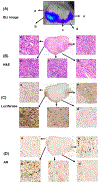
An example of tumor heterogeneity at gross appearance and histology level. (A) The magnified bioluminescence image of the tumor with arrows specifying where histopathological images were captured. Digital scans of (B) H&E of cross section of the tumor (scale bar, 3 mm) and IHC staining for (C) luciferase (scale bar, 3 mm) and (D) AR (scale bar, 4 mm). ‘a-e’ are higher magnification photomicrographs of specified regions of interests (scale bar, 100 µm).

References

    1. Huang Y, Cheng C, Zhang C, et al. Advances in prostate cancer research models: From transgenic mice to tumor xenografting models. Asian J Urol. 2016;3(2):64–74. - PMC - PubMed
    1. Cunningham D, You Z. In vitro and in vivo model systems used in prostate cancer research. J Biol Methods. 2015;2(1). - PMC - PubMed
    1. Valta M, Fagerlund K, Suominen M, Halleen J, Tuomela J. Importance of microenvironment in preclinical models of breast and prostate cancer. 2015.
    1. Zinn KR, Chaudhuri TR, Szafran AA, et al. Noninvasive Bioluminescence Imaging in Small Animals. Ilar j. 2008;49(1):103–115. - PMC - PubMed
    1. Ayers GD, McKinley ET, Zhao P, et al. Volume of preclinical xenograft tumors is more accurately assessed by ultrasound imaging than manual caliper measurements. J Ultrasound Med. 2010;29(6):891–901. - PMC - PubMed

Publication types

Substances