Review
. 2019 Aug 23;431(18):3472-3500.
doi: 10.1016/j.jmb.2019.04.002. Epub 2019 Apr 5.

β-Lactamases and β-Lactamase Inhibitors in the 21st Century

Affiliations
Review

β-Lactamases and β-Lactamase Inhibitors in the 21st Century

Catherine L Tooke et al. J Mol Biol. .

Abstract

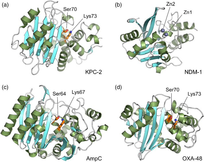
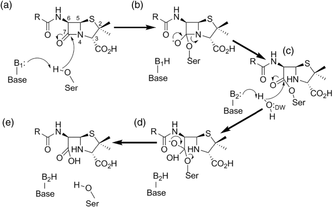
The β-lactams retain a central place in the antibacterial armamentarium. In Gram-negative bacteria, β-lactamase enzymes that hydrolyze the amide bond of the four-membered β-lactam ring are the primary resistance mechanism, with multiple enzymes disseminating on mobile genetic elements across opportunistic pathogens such as Enterobacteriaceae (e.g., Escherichia coli) and non-fermenting organisms (e.g., Pseudomonas aeruginosa). β-Lactamases divide into four classes; the active-site serine β-lactamases (classes A, C and D) and the zinc-dependent or metallo-β-lactamases (MBLs; class B). Here we review recent advances in mechanistic understanding of each class, focusing upon how growing numbers of crystal structures, in particular for β-lactam complexes, and methods such as neutron diffraction and molecular simulations, have improved understanding of the biochemistry of β-lactam breakdown. A second focus is β-lactamase interactions with carbapenems, as carbapenem-resistant bacteria are of grave clinical concern and carbapenem-hydrolyzing enzymes such as KPC (class A) NDM (class B) and OXA-48 (class D) are proliferating worldwide. An overview is provided of the changing landscape of β-lactamase inhibitors, exemplified by the introduction to the clinic of combinations of β-lactams with diazabicyclooctanone and cyclic boronate serine β-lactamase inhibitors, and of progress and strategies toward clinically useful MBL inhibitors. Despite the long history of β-lactamase research, we contend that issues including continuing unresolved questions around mechanism; opportunities afforded by new technologies such as serial femtosecond crystallography; the need for new inhibitors, particularly for MBLs; the likely impact of new β-lactam:inhibitor combinations and the continuing clinical importance of β-lactams mean that this remains a rewarding research area.

Keywords: antimicrobial resistance; carbapenemase; enzyme mechanism; metallo-β-lactamase; β-lactam.


Figures

Unlabelled Image
Graphical abstract
Fig. 1
Fig. 1
Structures of representative β-lactams and β-lactamase inhibitors. 1. Penicillin scaffold. 2. Cephalosporin scaffold. 3. (1-methyl) Carbapenem scaffold. 4. Hydrolyzed carbapenem (Δ2-pyrroline form). 5. Hydrolyzed carbapenem (Δ1-pyrroline form). 6. Monobactam scaffold. 7. Clavulanic acid. 8. Avibactam. 9. Relebactam. 10. Vaborbactam. 11. Bicyclic boronate.
Fig. 2
Fig. 2
Overall structure of representative β-lactamases from each class. Crystal structures of β-lactamases from classes A, B, C and D. Catalytic important residues of serine-β-lactamases (serine 64/70 and lysine 67/73, labeled) are colored orange, and the metallo-β-lactamase zinc ions are shown as gray spheres. (a) Class A KPC-2 (PDB 5ul8). (b) Class B NDM-1 (PDB 5zgy). (c) Class C AmpC (PDB 1ke4). (d) Class D OXA-48 (PDB 3hbr). Figure (and other structural Figures) generated with Pymol (www.pymol.org).
Fig. 3
Fig. 3
Mechanistic overview of serine β-lactamases. Figure shows hydrolysis of generic penicillin substrate. (a) General base B1 activates Ser for nucleophilic attack on the amide carbonyl carbon (C7) generating covalent acylenzyme (c) via tetrahedral oxyanionic acylation transition state (b). General base B2 activates incoming deacylating water molecule DW for nucleophilic attack on the acylenzyme carbonyl liberating penicilloate product (e) via tetrahedral deacylation transition state (d). For clarity, details of proton transfers to N4 and Ser are omitted. Note that the identities of bases B1 and B2 vary between β-lactamase classes.
Fig. 4
Fig. 4
(A) alternative acylation mechanisms for class A β-lactamases. (a) Apoprotein active site shows Lys73 protonated and Glu166 deprotonated. Ser70 may be activated for nucleophilic attack upon the β-lactam carbonyl by Glu166 via a water molecule (b). Alternatively, substrate binding reorganizes protonation in the active site such that neutral Lys73 activates Ser70 (c). The transition state (d) resolves to the acylenzyme intermediate (e) by protonation of the amide nitrogen from Ser130.
Fig. 5
Fig. 5
Carbapenem complexes of class A β-lactamases. Close-up views of class A: carbapenem complexes, carbapenem acylenzymes are covalently attached to Ser70. Note hydrogen bonding interactions of the acylenzyme carbonyl with the oxyanion hole formed by the backbone amides of residues 70 (nucleophilic Ser) and 237. (a) SHV-1:meropenem acylenzyme (PDB:2ZD8). Meropenem is modeled in two conformers with the carbonyl oxygen either pointing into the oxyanion hole (interacting with Ala237 and Ser70) or pointing away from the oxyanion hole (interacting with Ser130). In both models, the meropenem R group is not modeled due to disorder. (b) KPC-2:faropenem non-covalent product complex (5UJ4). (c) SFC-1:meropenem acylenzyme (4EV4). (d) GES-5:imipenem acylenzyme (4H8R). Important residues for catalysis and substrate binding (labeled) are represented as sticks, and the deacylating water is displayed as a red sphere. Distances (Å) are displayed as dashed lines.
Fig. 6
Fig. 6
Active-site architecture of class B metallo-β-lactamases and their carbapenem complexes. Close-up views of native metallo-β-lactamase active sites (left) and hydrolyzed carbapenem products (labeled) bound in the active sites (right). (a) B1 NDM-1 (PDBs 5zgx and 5ypk; left and right, respectively). (b) B1 VIM-1 (5n5g and 5n5i). (c) B3 SMB-1 (3vpe and 5b1u).
Fig. 7
Fig. 7
Possible mechanism of carbapenem hydrolysis by binuclear class B metallo-β-lactamases. (a) Substrate binding displaces Zn-bridging hydroxide to a terminal position (b) enabling attack upon the scissile carbonyl. (c) Anionic intermediate with negative charge delocalized around the pyrroline ring resolves either (d) by protonation at C2 by bulk water generating (e) the Δ1 pyrroline or (f) at N4 by incoming water at the bridging position generating (g) the Δ2-pyrroline product. (Note that as shown Zn-bound “apical” water is displaced by substrate; some proposals show this as moving to the bridging position on substrate binding.)
Fig. 8
Fig. 8
Proposed deacylation mechanisms for class C β-lactamases. Figure shows possible schemes for hydrolysis of generic cephalosporin acylenzyme. (a) Conjugate-base hypothesis where Lys67 deprotonates Tyr150 to activate deacylating water molecule. (b) Substrate-activated hypothesis whereby substrate N deprotonates deacylating water. Dashed lines denote hydrogen bonds.
Fig. 9
Fig. 9
Class C β-lactamase active sites. (a) AmpC:imipenem complex (PDB:1LL5), imipenem acylenzyme covalently attached to Ser64 (note the presence of putative deacylating water adjacent to Tyr150) and (b) ADC-68 active site (note the residues 320 and 321 in the putative C-loop associated with carbapenem turnover). Distances (in Å) displayed as dashed lines. Important residues are represented as sticks (labeled), and waters are shown as spheres.
Fig. 10
Fig. 10
Active sites of class D β-lactamases and carbapenem acylenzymes. (a) Native OXA-23 (PDB 4KOX; note the hydrophobic bridge between Phe110 and Met221 and carboxylated Lys82; deacylating water is shown as a red sphere). (b) OXA-23:meropenem acylenzyme [PDB 4JF4; note the carbapenem acylenzyme (yellow) in Δ1-pyrroline form]. (c) OXA-24/40:doripenem acylenzyme [PDB 3PAE; note the carbapenem acylenzyme (cyan) in Δ2-pyrroline form]. Carbapenem acylenzymes (b and c) shown as sticks covalently attached to Ser79. Distances (in Å) displayed as dashed lines. Note that, for consistency, residue numbering for all panels is as used by Smith etal. .
Fig. 11
Fig. 11
Interactions of β-lactamases with β-lactamase inhibitors. (a) Vaborbactam bound to class A CTX-M-15 (PDB 4xuz). (b) Avibactam bound to CTX-M-15 (PDB 4hbu). (c) A cyclobutanone bound to subclass B2 metallo-β-lactamase SPM-1 (PDB 5ndb). (d) A bicyclic boronate bound to subclass B1 metallo-β-lactamase VIM-2 (PDB 5fqc). Inhibitor molecules and the protein residues they interact, with are shown as sticks. Ligand interactions are shown as colored dashes.

References

    1. Fleming A. On the antibacterial action of cultures of a penicillium with special reference to their use in the isolation of B. influenzae. Br. J. Exp. Pathol. 1929;10:226–236. - PubMed
    1. Klein E.Y., Van Boeckel T.P., Martinez E.M., Pant S., Gandra S., Levin S.A., et al. Global increase and geographic convergence in antibiotic consumption between 2000 and 2015. Proc. Natl. Acad. Sci. U. S. A. 2018;115:E3463-E70. - PMC - PubMed
    1. Neu H.C. beta-Lactam antibiotics: structural relationships affecting in vitro activity and pharmacologic properties. Rev. Infect. Dis. 1986;8(Suppl. 3):S237–S259. - PubMed
    1. Brotzu G. 1948. Ricerche su di un nuovo antibiotico. Lavori dell'Istituto d'Igiene di Cagliari; pp. 4–18.
    1. Newton G.G., Abraham E.P. Degradation, structure and some derivatives of cephalosporin N. Biochem. J. 1954;58:103–111. - PMC - PubMed

Publication types

MeSH terms