Review
. 2019 Apr 6;11(4):794.
doi: 10.3390/nu11040794.

Analysis of Association between Vitamin D Deficiency and Insulin Resistance

Affiliations
Review

Analysis of Association between Vitamin D Deficiency and Insulin Resistance

Izabela Szymczak-Pajor et al. Nutrients. .

Abstract

Recent evidence revealed extra skeleton activity of vitamin D, including prevention from cardiometabolic diseases and cancer development as well as anti-inflammatory properties. It is worth noting that vitamin D deficiency is very common and may be associated with the pathogenesis of insulin-resistance-related diseases, including obesity and diabetes. This review aims to provide molecular mechanisms showing how vitamin D deficiency may be involved in the insulin resistance formation. The PUBMED database and published reference lists were searched to find studies published between 1980 and 2019. It was identified that molecular action of vitamin D is involved in maintaining the normal resting levels of ROS and Ca2+, not only in pancreatic β-cells, but also in insulin responsive tissues. Both genomic and non-genomic action of vitamin D is directed towards insulin signaling. Thereby, vitamin D reduces the extent of pathologies associated with insulin resistance such as oxidative stress and inflammation. More recently, it was also shown that vitamin D prevents epigenetic alterations associated with insulin resistance and diabetes. In conclusion, vitamin D deficiency is one of the factors accelerating insulin resistance formation. The results of basic and clinical research support beneficial action of vitamin D in the reduction of insulin resistance and related pathologies.

Keywords: insulin resistance; insulin-responsive tissues; oxidative stress; pancreatic β-cells dysfunction; sub-inflammation; vitamin D.


Conflict of interest statement

The authors declare no conflict of interest.

Figures
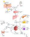
Figure 1
Figure 1
The regulation of synthesis and metabolism of vitamin D. Under ultraviolet radiation (UVB, 290–315 nm) action, 7-dehydrocholesterol in converted into previtamin D3 in the skin. In turn, previtamin D3 is immediately transformed into vitamin D3 as a result of heat-dependent process [10]. During excessive exposure to sun, previtamin D3 and vitamin D3 are broken down into inactive photoproducts to prevent vitamin D3 intoxication [11]. Both vitamin D2 and vitamin D3 derived from synthesis in the skin and a diet may be transported by vitamin D binding protein (VDBP) with the bloodstream or may be stored in adipocytes and then released to the circulation. The next step of vitamin D metabolism comprises two consecutive enzymatic hydroxylation reactions leading to vitamin D activation. The first step of vitamin D activation is the formation of 25(OH)D in the liver by vitamin D-25-hydroxylase, a cytochrome P450 enzyme, (mainly CYP2R1) [12]. The 1,25(OH)2D (calcitriol, the bioactive metabolite of vitamin D) forms as a result of 25(OH)D hydroxylation being performed by 25(OH)D-1α-hydroxylase (CYP27B1). This enzyme is present not only in the tubules of kidney, but also in numerous cells including macrophages, adipocytes, and the pancreatic β-cells [13,14,15,16]. The 1,25(OH)2D3 is able to induce its own degradation via the stimulation of 25(OH)D-24-hydroxylase (CYP24A1). CYP24A1 is an enzyme responsible for the degradation of both calcitriol and its precursor 25(OH)D to biological inactive metabolites, i.e., calcitroic acid excreted with the bile [11]. A low level of vitamin D and calcium stimulates parathyroid gland for the release of parathyroid hormone (PTH) and induction of CYP27B1 synthesis, resulting in elevated calcitriol activation [17]. The 1,25(OH)2D3 may reduce its own synthesis via negative feedback loop and decreases both synthesis and secretion of PTH. PTH is also capable of inhibition of CYP24A1 [18] and induction of skeletal fibroblast growth factor 23 (FGF-23) synthesis [19]. FGF-23 regulates the vitamin D homeostasis via inhibiting renal expression of CYP27B1 and stimulating expression of CYP24A1 which resulting in the reduction of calcitriol level in the serum [11]. formula image—stimulation, formula image—inhibition.
Figure 2
Figure 2
The insulin signaling pathway under physiological condition. Insulin action is initiated via its binding to insulin receptor (IR). The activation of IR contributes to the dimerization of the receptor and generation of the heterotetrameric form. Autophosphorylation of IR leads to the formation of numerous phosphotyrosine residues which are potential docking sites for the component of other signaling pathways [29]. The recruitment and phosphorylation of numerous substrate proteins, including insulin-receptor substrate (IRS) proteins, are allowed via multiple phosphotyrosines [30]. Phosphorylated IRSs activate and translocate phosphatidylinositol-3-kinase (PI3K) to the plasma membrane, and PI3K phosphorylates phosphatidylinositol 4,5-biphosphate (PIP2) to phosphatidylinositol-3,4,5-biphosphate (PIP3)—a key lipid signaling molecule. The level of PIP3 is under control of phosphatase and tensin homolog (PTEN) and SH2-containing inositol 5′-phosphatase-2 (SHIP2) that perform PIP3 dephosphorylation [28]. Insulin-mediated elevation of PIP3 level induces serine threonine kinase PDK1 (phosphoinositide-dependent protein kinase-1), thus leading to the phosphorylation and activation of protein kinase C (PKC ζ/λ) and protein kinase B (PKB also known as AKT). One of their actions is the translocation of glucose transporter 4 (GLUT4) to cell membrane and, in consequence, the elevation of glucose uptake [31]. AKT also stimulates synthesis of protein, glycogenesis, and lipogenesis, but represses lipolysis, glucogenolysis, gluconeogenesis, and proteolysis [28].
Figure 3
Figure 3
The attenuation of insulin signaling pathway in insulin resistance condition. Numerous protein kinases, i.e., IKK-β, JNK, PKC ζ/λ, PKC-θ, contribute to the phosphorylation of IRS that in turn attenuate insulin signaling. This state is presented in insulin resistance. formula image—attenuation.
Figure 4
Figure 4
Genomic mechanism of vitamin D action involved in the regulation of DNA demethylases genes expression. The 1,25(OH)2D3 binds to VDR, which in turn heterodimerizes with RXR. The formed 1,25(OH)2D3-VDR-RXR complex translocates to the nucleus where it binds to VDRE. As a result, the expression of vitamin D-dependent DNA demethylases, i.e., LSD1, LSD2, JMJD1A, and JMJD3, is upregulated. These enzymes prevent hypermethylation of promotor regions of numerous genes.
Figure 5
Figure 5
The 1,25(OH)2D3-mediated induction of adipocyte apoptosis. The 1,25(OH)2D3 stimulates both voltage-insensitive and voltage-dependent Ca2+ influx in mature adipocytes leading to the release of Ca2+ from ER stores via RyR and InsP3R. Increased intracellular Ca2+ level activates apoptosis via the Ca2+-dependent protease calpain contributing to the activation of the Ca2+/calpain-dependent caspase-12. Modified according to Abbas et al. [8]. formula image—activation.
Figure 6
Figure 6
The inhibitory effect of 1,25(OH)2D3 on inflammation. LPS- or TNF-α-stimulated receptors i.e., TLR, IL-6R activates P38MAPK- or NF-κB-dependent transcription of pro-inflammatory genes such as IL-1β, IL-6, TNF-α. The 1,25(OH)2D3 inhibits inflammation via suppression of IκBα phosphorylation and subsequent translocation of P38MAPK or NF-κB into the nucleus, leading to decreased expression of pro-inflammatory genes. formula image—activation, formula image—inhibition.

References

    1. Wang H., Chen W., Li D., Yin X., Zhang X., Olsen N., Zheng S.G. Vitamin D and Chronic Diseases. Aging Dis. 2017;8:346–353. doi: 10.14336/AD.2016.1021. - DOI - PMC - PubMed
    1. Tao S., Yuan Q., Mao L., Chen F.-L., Ji F., Cui Z.-H. Vitamin D deficiency causes insulin resistance by provoking oxidative stress in hepatocytes. Oncotarget. 2017;8:67605–67613. doi: 10.18632/oncotarget.18754. - DOI - PMC - PubMed
    1. Tabesh M., Azadbakht L., Faghihimani E., Tabesh M., Esmaillzadeh A. Effects of calcium-vitamin D co-supplementation on metabolic profiles in vitamin D insufficient people with type 2 diabetes: A randomised controlled clinical trial. Diabetologia. 2014;57:2038–2047. doi: 10.1007/s00125-014-3313-x. - DOI - PubMed
    1. Upreti V., Maitri V., Dhull P., Handa A., Prakash M.S., Behl A. Effect of oral vitamin D supplementation on glycemic control in patients with type 2 diabetes mellitus with coexisting hypovitaminosis D: A parellel group placebo controlled randomized controlled pilot study. Diabetes Metab. Syndr. 2018;12:509–512. doi: 10.1016/j.dsx.2018.03.008. - DOI - PubMed
    1. Barzegari M., Sarbakhsh P., Mobasseri M., Noshad H., Esfandiari A., Khodadadi B., Gargari B.P. The effects of vitamin D supplementation on lipid profiles and oxidative indices among diabetic nephropathy patients with marginal vitamin D status. Diabetes Metab. Syndr. 2019;13:542–547. doi: 10.1016/j.dsx.2018.11.008. - DOI - PubMed

MeSH terms