. 2019 Mar 5;11(3):419.
doi: 10.3390/polym11030419.

Microfluidic Based Fabrication and Characterization of Highly Porous Polymeric Microspheres

Affiliations

Microfluidic Based Fabrication and Characterization of Highly Porous Polymeric Microspheres

Benzion Amoyav et al. Polymers (Basel). .

Abstract

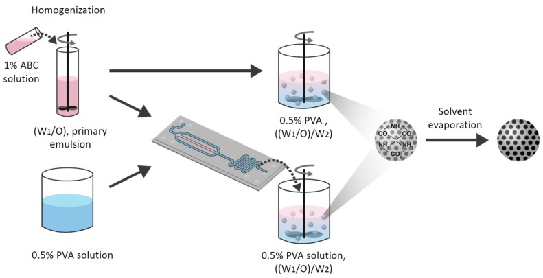
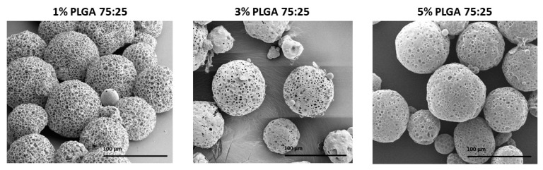
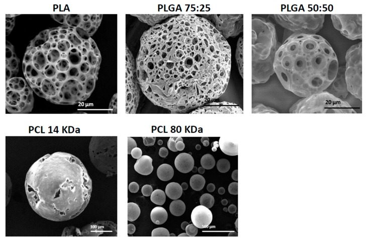
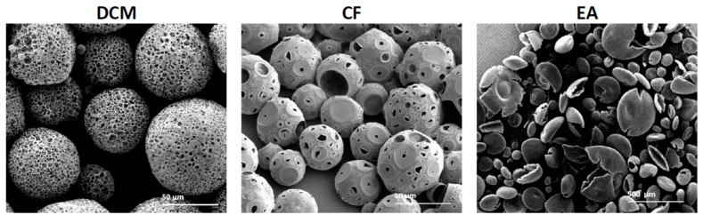
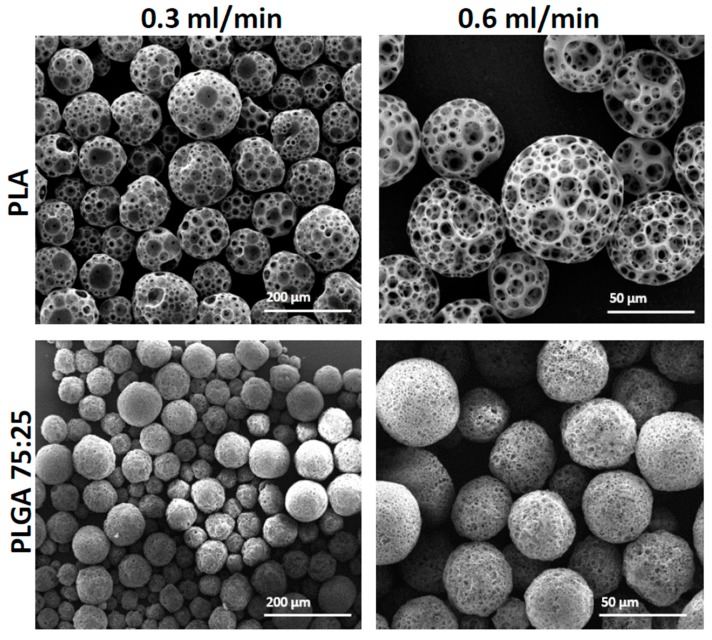
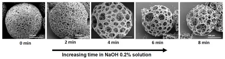
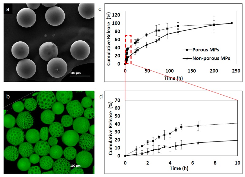
Polymeric porous particles are currently used for various applications in biotechnology, tissue engineering and pharmaceutical science, e.g., floating drug delivery systems and inhaled formulations. Particle shape and size depend on variable parameters; among them, polymer type and concentration, stirring speed, pH and type of solvent. In this study, porous poly(lactic-co-glycolic) acid (PLGA) and poly(d,l-lactide) (PLA) microspheres (MPs), with varying sizes and morphologies, were synthesized and optimized using both batch formulation and a flow-focusing microfluidic device. A well-established method of preparation utilizing solvent evaporation and the double emulsion technique was performed. Similar to other batch encapsulation methods, this technique is time and reagent consuming and consists of several steps. Hence, although porous structures provide tremendous opportunity in the design of new applications for tissue engineering and as improved controlled-release carriers, the synthesis of these particles with predefined properties remains challenging. We demonstrated the fabrication of porous MPs using a simple microfluidic device, compared to batch synthesis fabrication; and the effect of solvent, polymer concentration and type, post-hydrolysis treatment, on porosity degree. Moreover, a kinetic release study of fluorescent molecule was conducted for non-porous in comparison to porous particles. An overview of future prospects and the potential of these porous beads in this scientific area are discussed.

Keywords: PLA; PLGA; focused-flow; microfluidics; porosity; porous microspheres; tissue engineering.


Conflict of interest statement

The authors declare no conflict of interest. The funders had no role in the design of the study; in the collection, analyses, or interpretation of data; in the writing of the manuscript, or in the decision to publish the results.

Figures

Figure 1
Figure 1
Microfluidics system for preparation of porous microspheres (MPs) with varying properties. (a) Laboratory microfluidic flow system set up. Two syringe pumps precisely control the fluid volume and flow rate injected through the chip. Droplets are collected in the outlet into a stirred glass cup. (b) Schematic illustration of an enlarged junction in the focused-flow chip design used for MP synthesis. The flow through the orifice enables a controlled droplet break-up, which is required for yielding monodisperse MPs.
Figure 2
Figure 2
A schematic illustration of the preparation method of porous MPs using either “batch” or the microfluidic technique. Ammonium bicarbonate (ABC) 1% solution with polymer solution was homogenized to form a primary emulsion (W1/O). Then, the emulsion was introduced to either a vessel of 0.5% (w/v) polyvinyl alcohol (PVA) solution or into a microfluidic droplet generation chip. Finally, the secondary double emulsion ((W1/O)/W2) was stirred with an overhead propeller to ensure complete evaporation that forms porous solid MPs.
Figure 3
Figure 3
Scanning electron microscope (SEM) images of poly(lactic-co-glycolic) acid (PLGA) porous microsphere particles fabricated by batch synthesis with increasing PLGA 75:25 concentrations (% w/v), 1%, 3% and 5%. The scale bars are 100 μm.
Figure 4
Figure 4
SEM images showing the morphology of porous and non-porous microspheres obtained with different polymers and molecular weights using batch synthesis. PLA, PLGA 75:25, PLGA 50:50, PCL 14 kDa and PCL 80 kDa.
Figure 5
Figure 5
SEM images of PLGA (75:25) particles obtained by batch synthesis show the influence of organic solvent on microsphere formation and porosity with three solvents: dichloromethane (DCM), chloroform (CF) and ethyl acetate (EA).
Figure 6
Figure 6
SEM images of porous polymeric MPs fabricated with increased continuous phase flow by a microfluidic droplet generation device. PLA and PLGA 75:25 MPs were fabricated with either 0.3 mL/min or 0.6 mL/min 0.5% PVA continuous phase. The primary emulsion (W1/O) flow rate was kept constant at 0.05 mL/min.
Figure 7
Figure 7
SEM images of 3% (w/v) PLGA 75:25 porous microspheres obtained by the microfluidic technique at increasing soaking time in NaOH 0.2% solution. As immersion time increased, a more porous structure was obtained.
Figure 8
Figure 8
6-coumarin release kinetic profile of MPs fabricated with the microfluidic approach. Samples were collected at a number of points of time and fluorescence was measured by a plate reader. Empty particles were used as control references. (a) Non-porous MPs, (b) porous MPs, (c) cumulative release profile of 250 h and (d) cumulative release profile of the initial 10 h.

References

    1. Cai Y., Chen Y., Hong X., Liu Z., Yuan W. Porous microsphere and its applications. Int. J. Nanomed. 2013;8:1111–1120. - PMC - PubMed
    1. Gokmen M.T., Du Prez F. Progress in Polymer Science Porous polymer particles—A comprehensive guide to synthesis, characterization, functionalization and applications. Prog. Polym. Sci. 2012;37:365–405. doi: 10.1016/j.progpolymsci.2011.07.006. - DOI
    1. Zhou J., Fang T., Wen J., Shao Z., Dong J. Silk coating on poly (ε-caprolactone) microspheres for the delayed release of vancomycin. J. Microencapsul. 2011;28:99–107. doi: 10.3109/02652048.2010.534824. - DOI - PubMed
    1. Oh Y.J., Lee J., Seo J.Y., Rhim T., Kim S.H., Yoon H.J., Lee K.Y. Preparation of budesonide-loaded porous PLGA microparticles and their therapeutic efficacy in a murine asthma model. J. Control. Release. 2011;150:56–62. doi: 10.1016/j.jconrel.2010.11.001. - DOI - PubMed
    1. Shahriari D., Koffler J.Y., Tuszynski M.H. Hierarchically ordered porous and high-volume polycaprolactone microchannel scaffolds enhanced axon growth in transected spinal cords. Tissue Eng. Part A. 2017;23:415–425. doi: 10.1089/ten.tea.2016.0378. - DOI - PMC - PubMed

LinkOut - more resources