. 2018 Jul 16;10(7):776.
doi: 10.3390/polym10070776.

Confirmation of Bioinformatics Predictions of the Structural Domains in Honeybee Silk

Affiliations

Confirmation of Bioinformatics Predictions of the Structural Domains in Honeybee Silk

Andrea L Woodhead et al. Polymers (Basel). .

Abstract

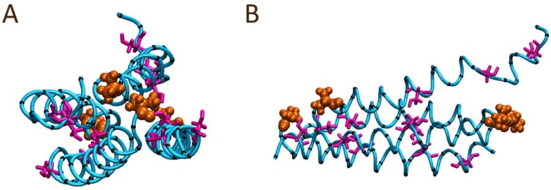
Honeybee larvae produce a silk made up of proteins in predominantly a coiled coil molecular structure. These proteins can be produced in recombinant systems, making them desirable templates for the design of advanced materials. However, the atomic level structure of these proteins is proving difficult to determine: firstly, because coiled coils are difficult to crystalize; and secondly, fibrous proteins crystalize as fibres rather than as discrete protein units. In this study, we synthesised peptides from the central structural domain, as well as the N- and C-terminal domains, of the honeybee silk. We used circular dichroism spectroscopy, infrared spectroscopy, and molecular dynamics to investigate the folding behaviour of the central domain peptides. We found that they folded as predicted by bioinformatics analysis, giving the protein engineer confidence in bioinformatics predictions to guide the design of new functionality into these protein templates. These results, along with the infrared structural analysis of the N- and C-terminal domain peptides and the comparison of peptide film properties with those of the full-length AmelF3 protein, provided significant insight into the structural elements required for honeybee silk protein to form into stable materials.

Keywords: bioinformatics protein folding prediction; cast film solubility; circular dichroism spectroscopy; coiled coil; infrared spectroscopy; molecular dynamics; protein design; protein engineering; protein materials; protein secondary structure.


Conflict of interest statement

The authors declare no conflict of interest.

Figures

Figure 1
Figure 1
Schematic representation of the locations of the peptides used in this study within the honeybee silk protein AmelF3.
Figure 2
Figure 2
Amide I regions of the FTIR spectra obtained from cast films of the four peptides derived from different regions of the honeybee silk protein, as shown in Figure 1. The dominant coiled coil (1649 cm−1) and β-sheet (1624 cm−1) bands are indicated. Deconvolution peaks are shown in Supplementary Information Figure S2 and the proportion of each structure is shown in Table 1.
Figure 3
Figure 3
Circular dichroism spectra for peptides AR28 (A) and AR99 (B) as a function of concentration.
Figure 4
Figure 4
A solution bound single AR28 peptide modeled in an initially extended conformation (A) and the final observed conformation (B) after 100 ns. The N-terminal alanine is shown in orange; the peptide backbone atoms are depicted as the cyan tube; Ser12, Ser20, and Ser26 sidechains are colored magenta; and all other sidechains and their respective α-carbons are shown in black.
Figure 5
Figure 5
Starting peptide alignment for the solution bound four AR28 peptide system modelled with a head-to-head alignment: (A) front and (B) perspective views. The N-terminal alanine is shown in orange; the peptide backbone atoms are depicted as the cyan tube; Ser12, Ser20, and Ser26 sidechains are colored magenta; and all other sidechains and their respective α-carbons are shown in black.
Figure 6
Figure 6
Final peptide alignment of the solution bound four AR28 peptide system modelled with an initial head-to-head alignment after 100 ns: (A) front and (B) side views. The N-terminal alanine is shown in orange; the peptide backbone atoms are depicted as the cyan tube; Ser12, Ser20, and Ser26 sidechains are colored magenta; and all other sidechain α-carbons are shown in black with all other sidechain atoms hidden for ease of viewing.
Figure 7
Figure 7
Starting peptide alignment for the solution bound four AR28 peptide system modelled with a head-to-tail alignment: (A) front and (B) perspective views. The N-terminal alanine is shown in orange; the peptide backbone atoms are depicted as the cyan tube; Ser12, Ser20, and Ser26 sidechains are colored magenta; and all other sidechains and their respective α-carbons are shown in black.
Figure 8
Figure 8
Final peptide alignment of the solution bound four AR28 peptide system modelled with an initial head-to-tail alignment after 100 ns: (A) front and (B) side views. The N-terminal alanine is shown in orange; the peptide backbone atoms are depicted as the cyan tube; Ser12, Ser20, and Ser26 sidechains are colored magenta; and all other sidechain α-carbons are shown in black with all other sidechain atoms hidden for ease of viewing.
Figure 9
Figure 9
Images of films cast from full-length honeybee silk protein (AmelF3), and peptides from different regions (Figure 1). Top panel, as cast films; Bottom panel, after aqueous methanol treatment, where materials cast from peptides have dissolved and are no longer obvious.

References

    1. Hepburn H.R., Kurstjens S.P. The combs of honeybees as composite materials. Apidologie. 1988;19:25–36. doi: 10.1051/apido:19880102. - DOI
    1. Silva-Zacarin E.C.M., Silva De Moraes R.L.M., Taboga S.R. Silk formation mechanisms in the larval salivary glands of Apis mellifera (Hymenoptera:Apidae) J. Biosci. 2003;28:753–764. doi: 10.1007/BF02708436. - DOI - PubMed
    1. Kameda T., Tamada Y. Variable-temperature 13C solid-state NMR study of the molecular structure of honeybee wax and silk. Int. J. Biol. Macromol. 2009;44:64–69. doi: 10.1016/j.ijbiomac.2008.10.004. - DOI - PubMed
    1. Sutherland T.D., Campbell P.M., Weisman S., Trueman H.E., Sriskantha A., Wanjura W.J., Haritos V.S. A highly divergent gene cluster in honey bees encodes a novel silk family. Genome Res. 2006;16:1414–1421. doi: 10.1101/gr.5052606. - DOI - PMC - PubMed
    1. Campbell P.M., Weisman S., Trueman H.E., Sriskantha A., Wanjura W.J., Haritos V.S. Cross-linking in the silks of bees, ants and hornets. Insect Biochem. Mol. Biol. 2014;48:40–50. doi: 10.1016/j.ibmb.2014.02.009. - DOI - PubMed