. 2018 Oct 11;10(10):1126.
doi: 10.3390/polym10101126.

Chromatin Loop Extrusion and Chromatin Unknotting

Affiliations

Chromatin Loop Extrusion and Chromatin Unknotting

Dusan Racko et al. Polymers (Basel). .

Abstract

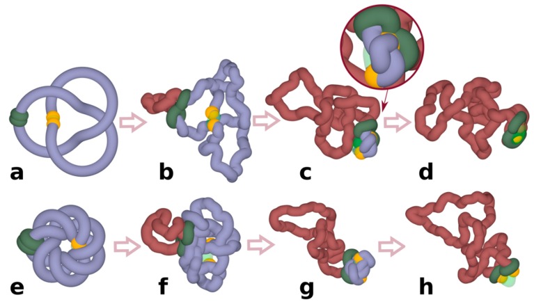
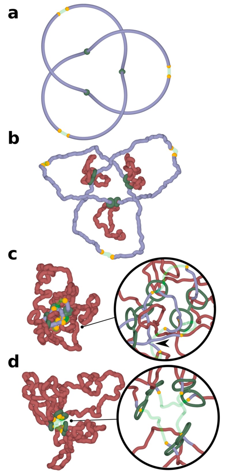
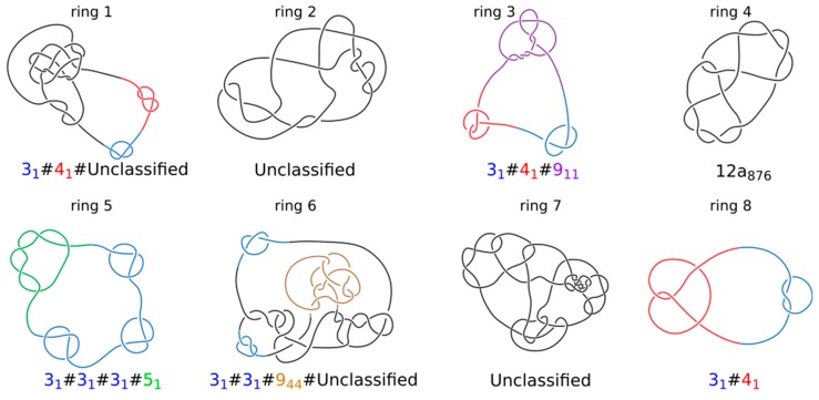
It has been a puzzle how decondensed interphase chromosomes remain essentially unknotted. The natural expectation is that in the presence of type II DNA topoisomerases that permit passages of double-stranded DNA regions through each other, all chromosomes should reach the state of topological equilibrium. The topological equilibrium in highly crowded interphase chromosomes forming chromosome territories would result in formation of highly knotted chromatin fibres. However, Chromosome Conformation Capture (3C) methods revealed that the decay of contact probabilities with the genomic distance in interphase chromosomes is practically the same as in the crumpled globule state that is formed when long polymers condense without formation of any knots. To remove knots from highly crowded chromatin, one would need an active process that should not only provide the energy to move the system from the state of topological equilibrium but also guide topoisomerase-mediated passages in such a way that knots would be efficiently unknotted instead of making the knots even more complex. We perform coarse-grained molecular dynamics simulations of the process of chromatin loop extrusion involving knotted and catenated chromatin fibres to check whether chromatin loop extrusion may be involved in active unknotting of chromatin fibres. Our simulations show that the process of chromatin loop extrusion is ideally suited to actively unknot, decatenate and demix chromatin fibres in interphase chromosomes.

Keywords: DNA knots; DNA topoisomerases; biopolymers; chromatin; chromatin loop extrusion; chromosomes; cohesin.


Conflict of interest statement

The authors declare no conflict of interest. The funders had no role in the design of the study; in the collection, analyses, or interpretation of data; in the writing of the manuscript, or in the decision to publish the results.

Figures

Figure 1
Figure 1
Chromatin loop extrusion unknots individual loop-forming TADs modelled as chromatin rings. (ad) Unknotting of a simple trefoil knot. (eh) Unknotting of a complex knot with 14 crossings. (c,d) show highly compressed knots being pushed towards a region with reduced excluded volume potential, which in our simulations mimic the action of Top2B, known to be present near TADs borders. Inset in (c) shows a close-up of a strongly confined knot, which is about to get unknotted by an intersegmental passage involving a region with reduced excluded volume potential. (d,h) show that at the end of simulations the knots are already unknotted. In our simulations, cohesin rings (green) are initially arbitrarily placed opposite to borders of modelled TADs. Once simulations are started, stacked rings of cohesin take the form of a quasi-planar handcuff and actively translocate in that form. Borders of TADs are recognizable here by yellow coloured beads that in our simulations stop the progression of cohesin rings and thus mimic the biological action of CTCF proteins located at borders of TADs. TAD: topologically associating domain.
Figure 2
Figure 2
Chromatin loop extrusion unknots a delocalized trefoil knot spanning a modelled chromosome portion containing three TADs. Three TADs, each with two cohesin rings forming handcuffs (green), beads mimicking action of CTCF (yellow) and TopII sites (green semi-transparent) are modelled as presented in Figure 1. (a) The starting configuration shows that the formed trefoil knot is not localized within an individual TAD, but diffuses over three TADs. (bd) Chromatin loop extrusion proceeds in all three TADs. The knot becomes excluded from the extruded chromatin portions and is concentrated in the chromatin portions with TopII sites of action, which are modelled as regions with reduced excluded volume potential. As the knot becomes more confined, it is more likely to interact with topoisomerases localized outside of TADs and is eventually unknotted there. Insets show close-ups of regions near TADs borders. For better visibility, the diameter of modelled chromatin fibres and cohesin handcuffs is decreased. In the inset shown in (c), the arrow indicates an unknotting passage involving a region with reduced excluded volume potential (semitransparent) and a chromatin portion that did not pass yet through cohesin rings (blue). In the inset shown in (d), the extrusion is nearly finished as cohesin rings are approaching sites with bound CTCF (yellow). The portion of chromatin, which will not be extruded further, is already free from knots.
Figure 3
Figure 3
Topology of knotted rings formed upon topological equilibration of 8 modelled chromatin rings. Knot types of all 8 rings forming a system presented in Figure 4. Alexander–Briggs notation of knots uses two numbers to indicate the knot type. The first number written in normal font indicates the minimal number of crossings of a given knot and the second number written as a subscript indicates the tabular position of this knot among the knots with the same minimal crossing number. So for example, 911 indicates a knot that in tables of knots is placed at 11th position among knots which minimal crossing number is 9. The # sign indicates that a knot is formed by a composition or merging together of simpler (prime) knots that are listed as its components. For prime knots with a large number of minimal crossings, e.g., 12, as is the case of ring 4, the knots are additionally divided into these with alternating pattern of crossings (indicated as a) and with nonalternating pattern of crossings (indicated as n). Standard tables of knots do not include knots with more than 12 crossings and we indicate these knots as unclassified.
Figure 4
Figure 4
Chromatin loop extrusion demixes topologically equilibrated chromatin loops. (a) A snapshot of topologically equilibrated system composed of 8 large chromatin circles (each shown in a different colour) that are knotted and catenated with each other. (b) A snapshot showing how the simulated system changes after each of the rings has undergone chromatin loop extrusion. (c) One of the 8 chromatin circles is shown together with its Voronoi envelope marking the interface of that chromatin circle with other circles. (d,e) Voronoi envelopes marking interfacial area between different chromatin circles before and after chromatin loop, respectively. (f) As chromatin loop extrusion progresses, the interfacial area between all modelled loops decreases. The snapshots present the confined chains together with their Voronoi envelopes at the moments when chromatin loop extrusion has started and finished, respectively. Red, encircled points indicate the interfacial area measures for the shown snapshots.

References

    1. Bates A.D., Maxwell A. DNA Topology. Oxford University Press; Oxford, UK: 2005. p. 198.
    1. Bjorkegren C., Baranello L. DNA Supercoiling, Topoisomerases, and Cohesin: Partners in Regulating Chromatin Architecture? Int. J. Mol. Sci. 2018;19 doi: 10.3390/ijms19030884. - DOI - PMC - PubMed
    1. Valdes A., Segura J., Dyson S., Martinez-Garcia B., Roca J. DNA knots occur in intracellular chromatin. Nucleic Acids Res. 2018;46:650–660. doi: 10.1093/nar/gkx1137. - DOI - PMC - PubMed
    1. O’Donnol D., Stasiak A., Buck D. Two convergent pathways of DNA knotting in replicating DNA molecules as revealed by theta-curve analysis. Nucleic Acids Res. 2018 doi: 10.1093/nar/gky559. - DOI - PMC - PubMed
    1. Portugal J., Rodriguez-Campos A. T7 RNA polymerase cannot transcribe through a highly knotted DNA template. Nucleic Acids Res. 1996;24:4890–4894. doi: 10.1093/nar/24.24.4890. - DOI - PMC - PubMed

LinkOut - more resources