

Humeral stress fracture in a female CrossFit athlete: a case report
- PMID: 30961567
- PMCID: PMC6454729
- DOI: 10.1186/s12891-019-2532-1
Humeral stress fracture in a female CrossFit athlete: a case report
Erratum in
-
Correction to: Humeral stress fracture in a female CrossFit athlete: a case report.BMC Musculoskelet Disord. 2019 Jun 18;20(1):289. doi: 10.1186/s12891-019-2674-1. BMC Musculoskelet Disord. 2019. PMID: 31208398 Free PMC article.
Abstract
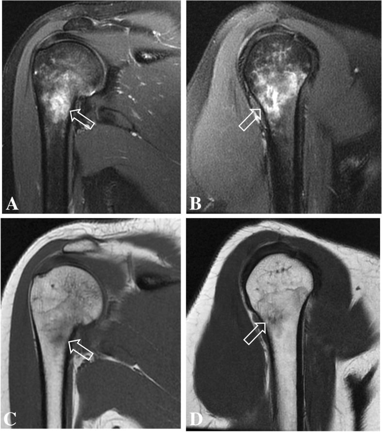
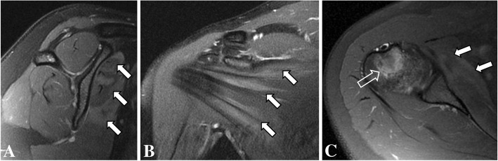
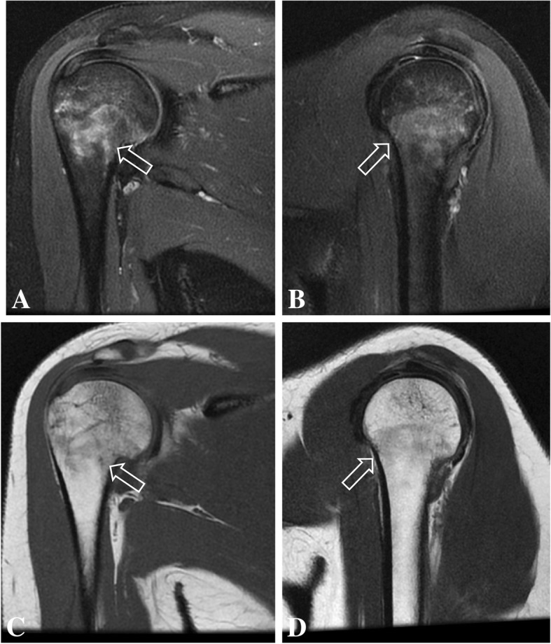
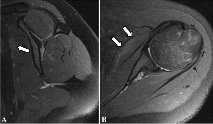
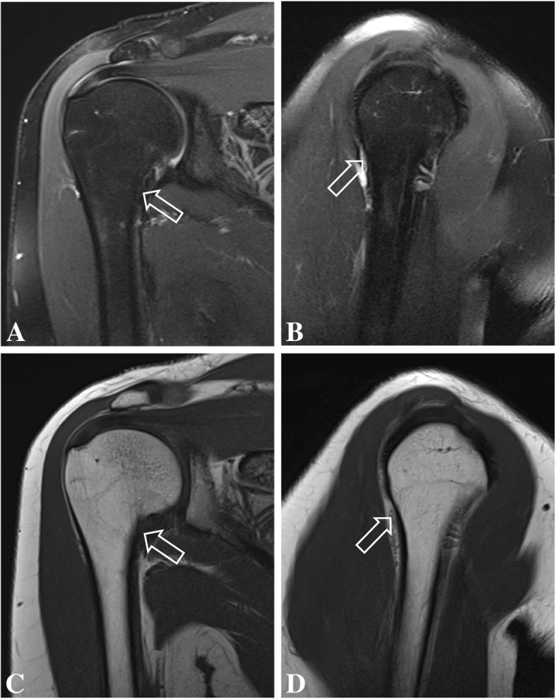
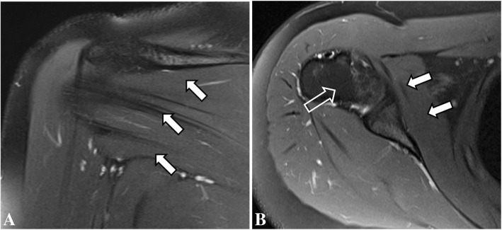
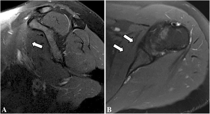
Background: Humeral stress fractures are rare injuries usually related to sports practice and joint overload without a direct trauma. A proximal humeral stress fracture has never been reported in a CrossFit athlete.
Case presentation: We report a stress fracture in the humerus of a 22-year-old woman after intense CrossFit training. Patient's previous medical history included amenorrhea and reduced Vitamin D levels. The patient was treated conservatively and resumed CrossFit training after she was advised not to until follow up imaging.
Conclusions: We present the MRI features of the case and emphasize the difficulties in diagnosis due to multiple possible causes of shoulder pain in a CrossFit athlete and by negative findings on early radiographs. Hormonal variations, Vitamin D insufficiency and the patient's attitude towards exercise were important factors that contributed for the stress injury after weight-lifting in CrossFit.
Keywords: CrossFit; Shoulder; Stress fracture.
Conflict of interest statement
Ethics approval and consent to participate
Not applicable.
Consent for publication
A written informed consent was obtained from the patient for publication of this case report and any accompanying images.
Competing interests
The authors declare that they have no competing interests.
Publisher’s Note
Springer Nature remains neutral with regard to jurisdictional claims in published maps and institutional affiliations.
Figures

Similar articles
-
Tarsal Navicular Stress Fracture in a Young Athlete: A Case Report.J Nippon Med Sch. 2019;86(2):122-125. doi: 10.1272/jnms.JNMS.2019_86-208. J Nippon Med Sch. 2019. PMID: 31130563
-
Unusual Stress Fracture in a CrossFit Athlete: A Case Report.JBJS Case Connect. 2021 Jan 14;11(1):e20.00135. doi: 10.2106/JBJS.CC.20.00135. JBJS Case Connect. 2021. PMID: 33502133
-
Case report: Longitudinal stress fracture of the humerus: imaging features and pitfalls.Clin Orthop Relat Res. 2009 Dec;467(12):3351-5. doi: 10.1007/s11999-009-0970-z. Epub 2009 Jul 9. Clin Orthop Relat Res. 2009. PMID: 19588209 Free PMC article.
-
Conservative Management of a Femoral Neck Stress Fracture in a Female Athlete. A Case Report and Review of the Literature.J Long Term Eff Med Implants. 2016;26(1):7-12. doi: 10.1615/JLongTermEffMedImplants.2016011991. J Long Term Eff Med Implants. 2016. PMID: 27649760 Review.
-
Taking a holistic approach to managing difficult stress fractures.J Orthop Surg Res. 2016 Sep 9;11(1):98. doi: 10.1186/s13018-016-0431-9. J Orthop Surg Res. 2016. PMID: 27608681 Free PMC article. Review.
Cited by
-
Vitamin D and Stress Fractures in Sport: Preventive and Therapeutic Measures-A Narrative Review.Medicina (Kaunas). 2021 Mar 1;57(3):223. doi: 10.3390/medicina57030223. Medicina (Kaunas). 2021. PMID: 33804459 Free PMC article. Review.
-
Rehabilitation After Surgical Treatment of Pectoralis Major Rupture in a CrossFit® Practitioner: A Case Report.Int J Sports Phys Ther. 2022 Jun 1;17(4):724-731. doi: 10.26603/001c.35720. eCollection 2022. Int J Sports Phys Ther. 2022. PMID: 35693859 Free PMC article.
-
Exploring the early diagnostic value of MRI for type I stress fractures: a retrospective analysis based on imaging manifestations.Front Surg. 2025 Mar 17;12:1333714. doi: 10.3389/fsurg.2025.1333714. eCollection 2025. Front Surg. 2025. PMID: 40166621 Free PMC article.
-
Humeral Fracture in a Young CrossFit Practitioner.Cureus. 2023 May 31;15(5):e39781. doi: 10.7759/cureus.39781. eCollection 2023 May. Cureus. 2023. PMID: 37398834 Free PMC article.
-
Correction to: Humeral stress fracture in a female CrossFit athlete: a case report.BMC Musculoskelet Disord. 2019 Jun 18;20(1):289. doi: 10.1186/s12891-019-2674-1. BMC Musculoskelet Disord. 2019. PMID: 31208398 Free PMC article.
References
-
- Horwitz BR, DiStefano V. Stress fracture of the humerus in a weight lifter. Orthopedics. 1995;18(2):185–187. - PubMed
-
- Schultz JT, Parker A, Curtis D, Daniel J, Huang H-H. International journal of exercise science: conference proceedings. 2016. The physiological and psychological benefits of CrossFit training–a pilot study; p. 14.
Publication types
MeSH terms
LinkOut - more resources
Full Text Sources
Medical
