Review
. 2019 Apr;5(4):245-262.
doi: 10.1016/j.trecan.2019.02.011. Epub 2019 Mar 22.

The Untranslated Regions of mRNAs in Cancer

Affiliations
Review

The Untranslated Regions of mRNAs in Cancer

Samantha L Schuster et al. Trends Cancer. 2019 Apr.

Abstract

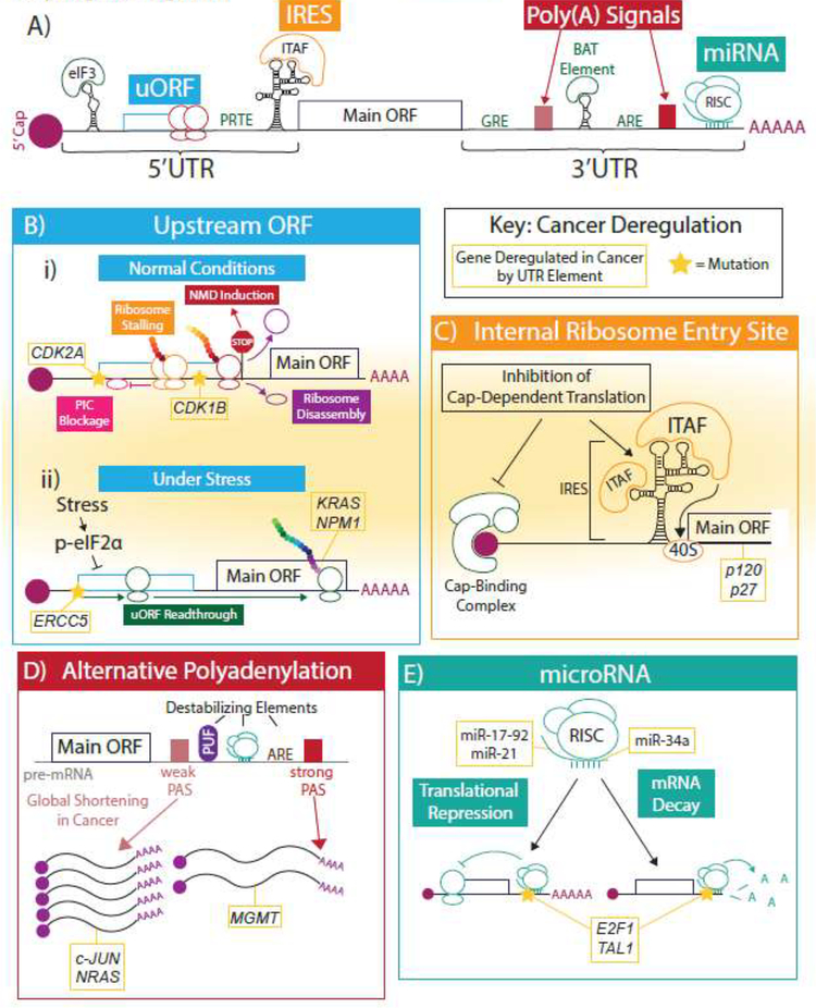
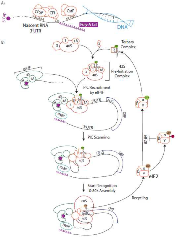
The 5' and 3' untranslated regions (UTRs) regulate crucial aspects of post-transcriptional gene regulation that are necessary for the maintenance of cellular homeostasis. When these processes go awry through mutation or misexpression of certain regulatory elements, the subsequent deregulation of oncogenic gene expression can drive or enhance cancer pathogenesis. Although the number of known cancer-related mutations in UTR regulatory elements has recently increased markedly as a result of advances in whole-genome sequencing, little is known about how the majority of these genetic aberrations contribute functionally to disease. In this review we explore the regulatory functions of UTRs, how they are co-opted in cancer, new technologies to interrogate cancerous UTRs, and potential therapeutic opportunities stemming from these regions.

Keywords: 3′UTR; 5′UTR; RNA metabolism; cancer; mRNA translation; somatic mutation; therapy; untranslated region.


Figures

Key Figure, Figure 1:
Key Figure, Figure 1:. 5’ and 3’UTR elements and how they are co-opted in cancer.
(A) The 5’ and 3’UTRs contain many regulatory elements. Here we show a theroretical mRNA schematic highlighting a few salient examples that have implications in cancer: uORFs, IRESs, poly(A) signals, miRNA binding sites, and several sequence- and structure-based motifs (in green). Throughout the figure, known examples of genes deregulated by each regulatory element in cancer are shown in gold boxes. Known mutations of regulatory elements in cancer are indicated with stars. (B) In normal conditions (i), upstream ORFs (uORFs) in the 5’UTR repress translation of the main downstream ORF through PIC blockage or stalling, nonsense-mediated decay, or PIC disassembly after translation of the uORF. Under stress (ii), where the translation machinery is downregulated, the PIC more often reads through the uORF and translates the downstream ORF more efficiently. (C) Internal ribosome entry sites (IRESs) are large structural elements in the 5’UTR that can, through the binding of IRES trans-acting factors (ITAFs), recruit the 40S to the start codon independent of cap-dependent translation. (D) Many genes contain multiple polyadenylation signals (PAS) in their 3’UTR that can give rise to differentially cleaved and polyadenylated transcripts. This results in the inclusion or exclusion of other, often destabilizing, cis-elements, affecting downstream gene expression. Often, use of proximal PASs increases gene expression and distal PASs decrease gene expression. (E) MicroRNAs (miRNAs) are small RNAs that integrate with the RNA-induced silencing complex (RISC) to mediate translational repression and/or mRNA decay of specific transcripts by binding target sequences, mostly in the 3’UTR.
Figure 2:
Figure 2:. Simplified schematic of mRNA processing and canonical cap-dependent translation.
A) Polyadenylation occurs co-transcriptionally in a two-step process of cleavage and adenylation. The polyadenylation signal consists of several upstream and downstream sequence motifs, which are recognized by the CPSF, CFI, and CstF complexes. B) Cap-dependent translation begins with the formation of the 43S pre-initiation complex (PIC) from the eukaryotic initiation factors eIF1, eIF1A, eIF3, eIF5, ternary complex (eIF2:GTP with meti-tRNA), and 40S small ribosomal subunit. The cap-binding eIF4F complex, consisting of eIF4E, eIF4G, and eIF4A, recruits the 43S PIC to the cap. The PIC then scans down the 5’UTR until recognition of a start codon. At the start codon, the 60S large ribosomal subunit joins and eIF2:GTP is hydrolyzed to eIF2:GDP. This must be recycled for subsequent rounds of initiation by the guanine exchange factor (GEF) eIF2B. Phosphorylation of the eIF2α subunit inhibits this GEF activity and therefore decreases availability of ternary complex and global translation levels.

Similar articles

Cited by

References

    1. Schwanhüusser B et al. (2011) Global quantification of mammalian gene expression control. Nature 473, 337–342 - PubMed
    1. Mularoni L et al. (2016) OncodriveFML: a general framework to identify coding and non-coding regions with cancer driver mutations. Genome Biol. 17, 128. - PMC - PubMed
    1. Weinhold N et al. (2014) Genome-wide analysis of noncoding regulatory mutations in cancer. Nat. Genet 46, 1160–1165 - PMC - PubMed
    1. Bailey MH et al. (2018) Comprehensive Characterization of Cancer Driver Genes and Mutations. Cell 173, 371–385.e18 - PMC - PubMed
    1. Network TCGAR (2014) Comprehensive molecular characterization of urothelial bladder carcinoma. Nature 507, 315–322 - PMC - PubMed

Publication types

LinkOut - more resources