. 2019 Jun 5;27(6):1087-1100.
doi: 10.1016/j.ymthe.2019.03.010. Epub 2019 Mar 19.

A Novel Endogenous Damage Signal, CSF-2, Activates Multiple Beneficial Functions of Adipose Tissue-Derived Mesenchymal Stem Cells

Affiliations

A Novel Endogenous Damage Signal, CSF-2, Activates Multiple Beneficial Functions of Adipose Tissue-Derived Mesenchymal Stem Cells

Se-Ra Park et al. Mol Ther. .

Abstract

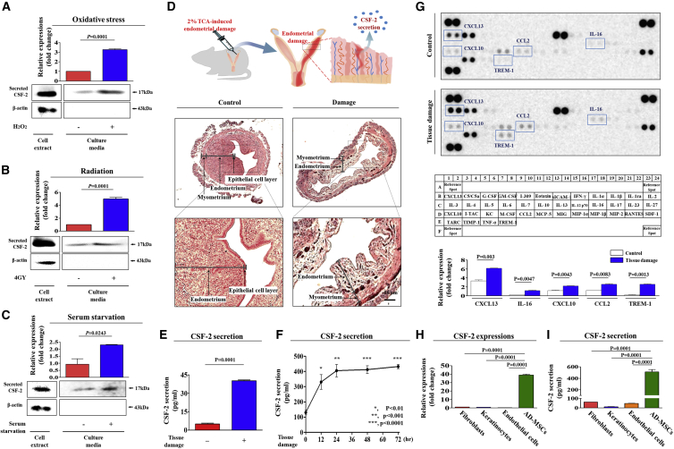
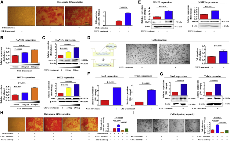
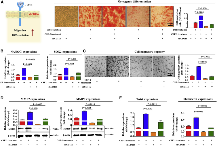
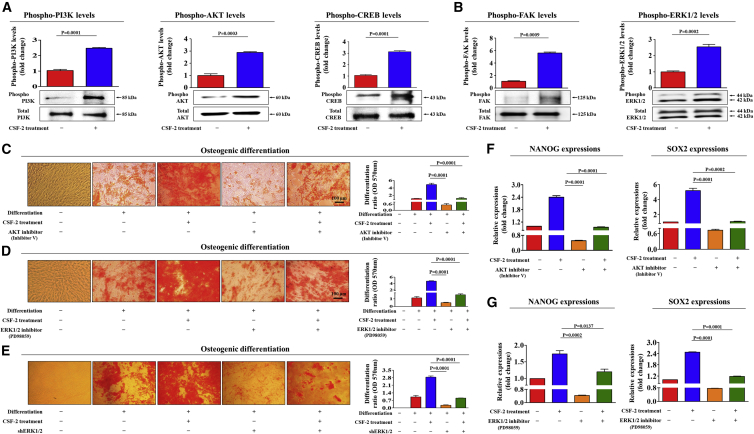
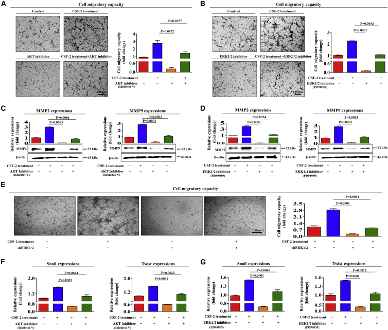
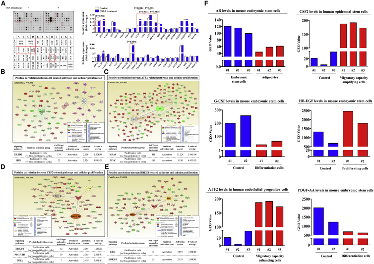
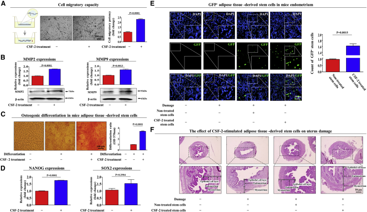
The major challenges of current mesenchymal stem cell (MSC)-based therapeutics are their low differentiation potential into specialized cell types and their homing ability to sites of injury. Therefore, many researchers have directed their efforts toward finding a novel stimulatory factor that can significantly enhance the therapeutic effects of MSCs. Colony-stimulating factor 2 (CSF-2) is previously known as a hematopoietic growth factor involved in the differentiation of various myeloid cells from hematopoietic progenitor cells. In addition to this canonical hematopoietic function, we identified for the first time that CSF-2 is actively secreted by stem cells, in response to various types of injuries, as an endogenous damage signal that promotes the therapeutic effects of MSCs by enhancing their multi-lineage differentiation and migratory capacities, possibly through its receptor CD116. Our results also revealed that CSF-2 exerts its stimulatory effects on MSCs via PI3K/Akt- and/or FAK/ERK1/2-signaling pathways. More importantly, we also found that MSCs stimulated with CSF-2 show markedly enhanced differentiation and migratory capacities and subsequent in vivo therapeutic effects in an endometrial ablation animal model. Collectively, our findings provide compelling evidence for a novel non-hematopoietic function of CSF-2 in promoting multiple beneficial functions of MSCs via a non-canonical mechanism as an endogenous damage signal.

Keywords: Akt; CSF-2; ERK1/2; MSCs; differentiation; growth; migration; stemness.


Figures

None
Graphical abstract
Figure 1
Figure 1
CSF-2 Is Actively Secreted by Stem Cells in Response to Various Injury Signals In Vitro and In Vivo (A) Mesenchymal stem cells (MSCs) were incubated in standard culture medium with or without H2O2 (10 mM) for 30 min, after which the medium was replaced with serum-free medium, and the cells were cultured for 48 h. (B) MSCs were exposed to acute irradiation at a dose of 4 Gy (X-ray), after which the medium was replaced with serum-free medium, and the cells were cultured for 48 h. (C) MSCs were cultured with or without serum for 48 h. (D) The 2% TCA treatment (150 μL, administered directly into the uterine horn) produced significant histological uterine endometrial ablation. (E) Increased CSF-2 concentrations by TCA treatment in the serum samples were also detected by ELISA. (F) TCA-induced acute endometrial ablation resulted in a clearly time-dependent manner, with a peak release 24 h after injury. (G) Cytokine/growth factor assay was performed using damaged and non-damaged samples. The membrane was printed with antibodies for 40 growth factors, cytokines, and receptors, with four positive and four negative controls in the upper and lower left corners. Five growth factors or related proteins (CXCL13, IL-16, CXCL10, CCL2, and TREM-1) were markedly enriched in the damaged groups compared with those in the control groups. The expression and secretion levels of CSF-2 in multiple cell types, such as fibroblasts, keratinocytes, endothelial cells, and MSCs, were detected by qPCR (H) and ELISA (I), respectively. β-actin was used as the internal control. The results represent the mean ± SD from three independent experiments.
Figure 2
Figure 2
CSF-2 Promotes the Differentiation and Migratory Capacities of MSCs In Vitro Confluent MSCs were cultured in osteogenic medium with or without CSF-2 (300 ng/mL). The effects of CSF-2 on osteoblast differentiation were determined by alizarin red staining. (A) The relative quantification of calcium mineral content was determined by measuring the absorbance at 570 nm. Real-time PCR (B) and western blotting (C) results demonstrating changes in the expression of the stem cell markers NANOG and SOX2 after CSF-2 treatment for 24 h. (D) MSCs were treated with CSF-2 for 24 h, after which the effect of CSF-2 on MSC migration ability was evaluated using the transwell migration assay. CSF-2 treatment significantly increased MSC migration across the membrane compared with the negative control. (E) The relative expression levels of key positive regulators of cell migration (MMP-2/9) were assessed using western blotting. Real-time PCR (F) and western blotting (G) results demonstrating changes in the expression of various EMT (epithelial-to-mesenchymal transition) markers, such as snail, and twist. MSCs were treated with 300 ng/mL CSF-2 alone or were concomitantly treated with neutralizing antibody targeting CSF-2; subsequent changes in osteoblast differentiation (H) and migratory capacity (I) were measured with alizarin red staining and transwell assay, respectively. β-actin was used as the internal control. The results represent the mean ± SD from three independent experiments.
Figure 3
Figure 3
CSF-2 Stimulates Multiple Beneficial Functions of MSCs through CD116 MSCs were treated with 300 ng/mL CSF-2 alone or were concomitantly transfected with shRNA targeting CD116; subsequent changes in osteoblast differentiation (A) and in the expression of pluripotency-associated factors NANOG and SOX2 (B) were measured with alizarin red staining and real-time PCR, respectively. The relative quantification of calcium mineral content was determined by measuring the absorbance at 570 nm. The inhibitory effect of CD116 knockdown on CSF-2-induced changes in the migratory capacity were measured by the transwell assay (C) and western blotting for MMP-2 and MMP-9 (D). (E) The attenuating effects of CD116 depletion on CSF-2-induced expression of fibronectin and twist in MSCs were measured by real-time PCR. β-actin was used as the internal control. The data represent the mean ± SD from three independent experiments.
Figure 4
Figure 4
CSF-2-Induced Stimulatory Effects on Stem Cells Are Mediated through PI3K/Akt and/or FAK/ERK1/2 Signaling (A and B) MSCs were stimulated for 10 min with or without CSF-2 (300 ng/mL). The cells were then lysed, and the protein contents were analyzed by western blotting using antibodies targeting the phosphorylated forms of PI3K, AKT, and CREB (A) and FAK and ERK1/2 (B). The phosphorylation levels of these signaling molecules were significantly increased in cells treated with CSF-2. (C and D) MSCs were pretreated with inhibitor V (C; 10 μM) or PD98059 (D; 20 μM) for 1 h prior to the additional treatment with 300 ng/mL CSF-2; subsequent changes in osteoblast differentiation were determined by alizarin red staining. (E) MSCs were treated with 300 ng/mL CSF-2 alone or were concomitantly transfected with shRNA targeting ERK1/2; subsequent changes in osteoblast differentiation were measured with alizarin red staining. (F and G) The expression of pluripotency-associated factors NANOG and SOX2 were determined by real-time PCR. The relative quantification of calcium mineral content was determined by measuring the absorbance at 570 nm. β-actin was used as the internal control. The data represent the mean ± SD from three independent experiments.
Figure 5
Figure 5
Inhibition of Akt or ERK1/2 with Specific Inhibitors Attenuated the CSF-2-Induced Migratory Capacity of MSCs (A–D) MSCs were pretreated with inhibitor V (A and C, 10 μM) or PD98059 (B and D, 20 μM) for 1 h prior to an additional 48-h treatment with 300 ng/mL CSF-2; subsequent changes in migratory capacity were measured via the transwell assay (A and B) and western blotting for MMP-2 and MMP-9 (C and D). (E) MSCs were treated with 300 ng/mL CSF-2 alone or were concomitantly transfected with shRNA targeting ERK1/2; subsequent changes in the migratory capacity were measured by the transwell assay. (F and G) MSCs were pretreated with inhibitor V (F, 10 μM) or PD98059 (G, 20 μM) for 1 h prior to an additional 48-h treatment with 300 ng/mL CSF-2; subsequent changes in the expressions of multiple EMT markers, such as snail and twist (F and G), were measured by real-time PCR. β-actin was used as the internal control. The data represent the mean ± SD from three independent experiments.
Figure 6
Figure 6
CSF-2-Induced Multiple Growth Factors Are Associated with PI3K/Akt and/or FAK/ERK 1/2 Signaling Human growth factor antibody array analysis was performed using CSF-2-treated and control samples. The membrane was printed with antibodies for 40 growth factors, cytokines, and receptors, with four positive and four negative controls in the upper and lower left corners. (A) Six growth factors or related proteins (AR, G-CSF, GDNF, GM-CSF, HB-EGF, and PDGF) were markedly enriched in the CSF-2-treated groups compared with those in the control groups. (B–E) Differentially expressed genes from proliferative cells and nonproliferative cells were applied to ingenuity pathway analysis (IPA) software (https://www.qiagenbioinformatics.com/) to predict the activation state (either activated or inhibited) of six growth factors and their related signaling pathways. Big data were analyzed using the Seiber dataset (GEO: GSE63074, GSE62564, GSE63074, and GSE28878) from R2: Genomics Analysis and Visualization Platform (https://hgserver1.amc.nl/cgi-bin/r2/main.cgi). The gene datasets were filtered by the expression profiles of six growth factors in proliferative versus nonproliferative cells. (F) The GEO database (https://www.ncbi.nlm.nih.gov/geo/) was analyzed to further verify the expression levels of these growth factors in various aspects of stem cell functions. The results represent the means ± SD from three independent experiments.
Figure 7
Figure 7
CSF-2 Improves the Therapeutic Potential of MSCs by Stimulating Differentiation and Migratory Capacities in a TCA-Induced Endometrial Ablation Animal Model Schematic representation of the experimental protocol as described in the Materials and Methods. Mice were treated daily for 10 days with CSF-2 (0.5 mg/kg, intravenously) or vehicle (PBS). Stem cells were isolated from mouse adipose tissue, and the changes in migratory capacity were measured via the transwell assay (A) and western blotting for MMP-2 and MMP-9 (B). The changes in osteoblast differentiation were determined by alizarin red staining. (C) The relative quantification of calcium mineral content was performed by measuring the absorbance at 570 nm. (D) Real-time PCR results showed the changes in the expression of the mouse stem cell markers NANOG and SOX2 after CSF-2 treatment in vivo. (E) The 2% TCA treatment (150 μL, administered directly into the uterine horn) produced significant histological uterine endometrial ablation compared with the vehicle (PBS) control. CSF-2-treated or non-treated MSCs (1 × 106 cells) were labeled with GFP and injected intravenously into the tail veins of 7-week-old immunodeficient NSG mice with acute TCA-induced endometrial ablation. The mice were sacrificed 7 days after the GFP-labeled MSCs were injected. Green fluorescent images of consecutive sections revealed the presence of GFP-labeled cells. (F) Uterine endometrial tissue was collected and subjected to H&E staining. TCA-induced loss of the endometrial functional layer with degenerative changes was significantly more relieved by the transplantation of CSF-2-pretreated MSCs. β-actin was used as the internal control. The data represent the mean ± SD from eight independent experiments.

Similar articles

Cited by

References

    1. Metcalf D. The molecular control of cell division, differentiation commitment and maturation in haemopoietic cells. Nature. 1989;339:27–30. - PubMed
    1. Ha Y., Kim Y.S., Cho J.M., Yoon S.H., Park S.R., Yoon D.H., Kim E.Y., Park H.C. Role of granulocyte-macrophage colony-stimulating factor in preventing apoptosis and improving functional outcome in experimental spinal cord contusion injury. J. Neurosurg. Spine. 2005;2:55–61. - PubMed
    1. Egea L., Hirata Y., Kagnoff M.F. GM-CSF: a role in immune and inflammatory reactions in the intestine. Expert Rev. Gastroenterol. Hepatol. 2010;4:723–731. - PMC - PubMed
    1. Pastore S., Fanales-Belasio E., Albanesi C., Chinni L.M., Giannetti A., Girolomoni G. Granulocyte macrophage colony-stimulating factor is overproduced by keratinocytes in atopic dermatitis. Implications for sustained dendritic cell activation in the skin. J. Clin. Invest. 1997;99:3009–3017. - PMC - PubMed
    1. Egea L., McAllister C.S., Lakhdari O., Minev I., Shenouda S., Kagnoff M.F. GM-CSF produced by nonhematopoietic cells is required for early epithelial cell proliferation and repair of injured colonic mucosa. J. Immunol. 2013;190:1702–1713. - PMC - PubMed

Publication types

MeSH terms

Substances