Observational Study
. 2019 May 6;216(5):1154-1169.
doi: 10.1084/jem.20190185. Epub 2019 Apr 8.

Longitudinal profiling of human blood transcriptome in healthy and lupus pregnancy

Affiliations
Observational Study

Longitudinal profiling of human blood transcriptome in healthy and lupus pregnancy

Seunghee Hong et al. J Exp Med. .

Abstract

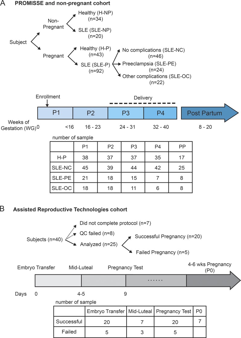
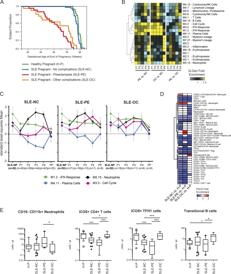
Systemic lupus erythematosus carries an increased risk of pregnancy complications, including preeclampsia and fetal adverse outcomes. To identify the underlying molecular mechanisms, we longitudinally profiled the blood transcriptome of 92 lupus patients and 43 healthy women during pregnancy and postpartum and performed multicolor flow cytometry in a subset of them. We also profiled 25 healthy women undergoing assisted reproductive technology to monitor transcriptional changes around embryo implantation. Sustained down-regulation of multiple immune signatures, including interferon and plasma cells, was observed during healthy pregnancy. These changes appeared early after embryo implantation and were mirrored in uncomplicated lupus pregnancies. Patients with preeclampsia displayed early up-regulation of neutrophil signatures that correlated with expansion of immature neutrophils. Lupus pregnancies with fetal complications carried the highest interferon and plasma cell signatures as well as activated CD4+ T cell counts. Thus, blood immunomonitoring reveals that both healthy and uncomplicated lupus pregnancies exhibit early and sustained transcriptional modulation of lupus-related signatures, and a lack thereof associates with adverse outcomes.


Figures

None
Graphical abstract
Figure 1.
Figure 1.
PROMISSE study design. (A) 92 SLE pregnant (SLE-P), 43 H-P, 20 SLE-NP, and 34 H-NP females were recruited. Pregnant SLE patients included NC (n = 46), PE (n = 24), and OC (n = 22). Blood was drawn in PAX gene tubes for microarray at specific time points (P1: <16 WG; P2: 16–23 WG; P3: 24–31 WG; P4: 32–40 WG, and between 8 and 20 wk PP). (B) Blood from 25 healthy women who underwent ART was analyzed for before and after embryo transfer by RNA-seq. Pregnancy rate for the cohort was 80% (20/25). Samples were taken on the day of the embryo transfer, midluteal, day of the pregnancy test, and P0 (4–6 WG).
Figure 2.
Figure 2.
Global blood transcriptome alterations during healthy pregnancy. (A) Hierarchical clustering of the 9,576 DETs between H-P and H-NP individuals. The list of DETs was obtained from the union of all transcripts identified as significantly modulated by the model through contrasts considering each pregnancy time point vs. H-NP (FDR-adjusted P ≤ 0.05). The line chart represents the molecular distance to health (MDTH) for individual samples, calculated as the sum of absolute FCs ≥2 for each transcript. (B) Principal component analysis based on the 9,576 transcripts in A. Dots represent individual samples and color coded by pregnancy group. (C) Blood module fingerprint of healthy pregnancy (P1 to P4, combined) in M1–7. Each dot on the grid represents a module, either overexpressed (red) or underexpressed (blue) as compared with H-NP controls. The intensity of the dots represents the percentage of transcripts from the modules that pass the over- or underexpression threshold. (D) Blood module functional map. PC, principal component; var, variance.
Figure 3.
Figure 3.
Increased neutrophil and decreased IFN and plasma cell signatures during healthy pregnancy. (A) Heatmap representing the Q-Gen fold enrichment for all pregnancy time points combined (H-P, P1–P4) and each individual time point vs. H-NP. Blood modules were used as gene sets. Modules modulated in any comparisons with FDR-adjusted P ≤ 0.05 and ≥30% FC were selected. (B) Line charts representing the standard least-squares mean for inflammation, erythropoiesis, neutrophils, IFN response, and plasma cell modules. Data are normalized to H-NP controls. (C) Box plots representing the absolute numbers of circulating neutrophils (granular CD14 CD66b+) and transitional B cells (CD20+ IgD+ CD24+ CD38++) during H-P and at PP in flow cytometry analysis. Error bars represent standard deviation. ***, P < 0.001.
Figure 4.
Figure 4.
Decreased IFN signature during successful embryo implantation. (A) Heatmap representing the Q-Gen fold enrichment for each time point vs. embryo transfer in failed (n = 5) and successful (n = 20) pregnancies. Blood modules were used as gene sets, and modules changed with nominal P ≤ 0.05 and ≥30% FC were selected and represented. (B) Line chart of the Q-Gen fold enrichment for IFN (M1.2) and erythropoiesis (M2.3) modules for successful or failed pregnant subjects. Significantly modulated time points compared with their own embryo transfer are indicated with asterisks (P ≤ 0.05). (C) DETs in successful vs. failed pregnancy at pregnancy test (nominal P ≤ 0.05, FC >1.25) are represented. Genes either in IFN modules or Interferome v2.01 (Rusinova et al., 2013) are highlighted in red.
Figure 5.
Figure 5.
Blood transcriptome alterations in noncomplicated SLE pregnancy. (A) Blood module fingerprints of SLE-NP (n = 20) vs. H-NP (n = 34), SLE pregnant with NC (SLE-NC; n = 46) vs. H-P (n = 43) and SLE-NC vs. SLE-NP. Each dot on the grid represents a module, either overexpressed (red) or underexpressed (blue) as compared with H-NP controls. The intensity of the dots represents the percentage of transcripts from the modules that pass the over- or underexpression threshold. (B) Hierarchical cluster of 3,138 DETs between SLE-NC and H-P at any time point. Transcript statistics across all comparisons are highlighted in the significance matrix on the right. The number of significant transcripts for each comparison (FDR-adjusted P ≤ 0.05) is displayed as a bar chart above the matrix. (C) Heatmap representing the blood module Q-Gen fold enrichment for each time point in SLE-NC vs. SLE-NP. Modules with FDR-adjusted P ≤ 0.05 and 30% FC in at least one condition were selected. (D) Line chart of the standard least-squares mean for inflammation, erythropoiesis, neutrophils, IFN response, and plasma cell modules in SLE-NP and SLE-NC normalized to H-NP controls throughout pregnancy. Sig, significant.
Figure 6.
Figure 6.
Blood transcriptome alteration in complicated SLE pregnancy. (A) Kaplan-Meyer curve representing gestational age at the end of pregnancy in H-P (n = 43), SLE-NC (n = 46), SLE-PE (n = 24), and SLE-OC (n = 22) cohorts. (B) Heatmap representing the blood module Q-Gen fold enrichment for indicated comparisons at specific time points. Modules with FDR-adjusted P ≤ 0.05 and ≥30% FC in at least one condition were selected. (C) Line chart depicting standard least-squares mean for IFN responses, plasma cells, neutrophils, and cell cycle in SLE-NC, PE, and OC. All values are normalized to H-NP controls. (D) Heatmap representing the normalized fold enrichment in FACS-analyzed leukocyte subsets at P1 and P2 combined. Red represents a statistically significant increase (FDR-adjusted P ≤ 0.05), blue a statistically significant decrease (FDR-adjusted P ≤ 0.05), and white a non–statistically significant change. Asterisks indicate data obtained from complete blood counts. (E) Box plot of flow cytometry analysis for a subset of patients (27 SLE-NC, 5 SLE-PE, and 4 SLE-OC) at P1 and P2 combined for immature neutrophils (granular fraction, CD14 CD16 CD11b), ICOS+ CD4+ T cells, ICOS+ CD4+ XCR5+ Tfh cells, and transitional B cells (CD20+ CD38+ CD24+ CD27). ****, P < 0.0001.
Figure 7.
Figure 7.
Identification of a predictive transcriptional signature of SLE PE in the first trimester of pregnancy. (A) Accuracy of different models predicting PE (n = 21) vs. NC (n = 45) at P1 using laboratory/clinical variables (gray) or gene expression only (black) or laboratory/clinical variables and gene expression combined (brown). (B) Receiver operator characteristic (ROC) curves of six models predicting PE vs. NC at P1 using laboratory/clinical variables only (gray), gene expression only (black), or both combined (brown). Area-under-the curve (AUC) measures for each model are displayed in the bottom right corner. (C) Top 58 genes selected by 5 out of 6 models at least once during cross-validation from A. Scaled expression of the genes in PE and NC are shown. GBM, gradient boosting machine; GLMNET, generalized linear model via penalized maximum likelihood; kernal PLS, kernel partial least squares; PAM, prediction analysis for microarrays; plsRglm, partial least-squares regression for generalized linear models; XGB, eXtreme gradient boosting.

Comment in

  • When pregnancy tames the wolf.
    Niewold TB, Mehta-Lee S. Niewold TB, et al. J Exp Med. 2019 May 6;216(5):1012-1013. doi: 10.1084/jem.20190378. Epub 2019 Apr 8. J Exp Med. 2019. PMID: 30962247 Free PMC article.

Similar articles

Cited by

References

    1. Aboussahoud W., Bruce C., Elliott S., and Fazeli A.. 2010. Activation of Toll-like receptor 5 decreases the attachment of human trophoblast cells to endometrial cells in vitro. Hum. Reprod. 25:2217–2228. 10.1093/humrep/deq185 - DOI - PubMed
    1. ACOG Committee on Practice Bulletins–Obstetrics 2002. ACOG practice bulletin. Diagnosis and management of preeclampsia and eclampsia. Number 33, January 2002. Obstet. Gynecol. 99:159–167. - PubMed
    1. Aghaeepour N., Ganio E.A., Mcilwain D., Tsai A.S., Tingle M., Van Gassen S., Gaudilliere D.K., Baca Q., McNeil L., Okada R., et al. . 2017. An immune clock of human pregnancy. Sci. Immunol. 2:eaan2946. 10.1126/sciimmunol.aan2946 - DOI - PMC - PubMed
    1. Allantaz F., Chaussabel D., Stichweh D., Bennett L., Allman W., Mejias A., Ardura M., Chung W., Smith E., Wise C., et al. . 2007. Blood leukocyte microarrays to diagnose systemic onset juvenile idiopathic arthritis and follow the response to IL-1 blockade [published correction appears in J Exp Med. 2009;206(10):2299]. J. Exp. Med. 204:2131–2144. 10.1084/jem.20070070 - DOI - PMC - PubMed
    1. Andrade D., Kim M., Blanco L.P., Karumanchi S.A., Koo G.C., Redecha P., Kirou K., Alvarez A.M., Mulla M.J., Crow M.K., et al. . 2015. Interferon-α and angiogenic dysregulation in pregnant lupus patients who develop preeclampsia. Arthritis Rheumatol. 67:977–987. 10.1002/art.39029 - DOI - PMC - PubMed

Publication types