. 2019 Jun 21;201(14):e00766-18.
doi: 10.1128/JB.00766-18. Print 2019 Jul 15.

ϕSa3mw Prophage as a Molecular Regulatory Switch of Staphylococcus aureus β-Toxin Production

Affiliations

ϕSa3mw Prophage as a Molecular Regulatory Switch of Staphylococcus aureus β-Toxin Production

Phuong M Tran et al. J Bacteriol. .

Abstract

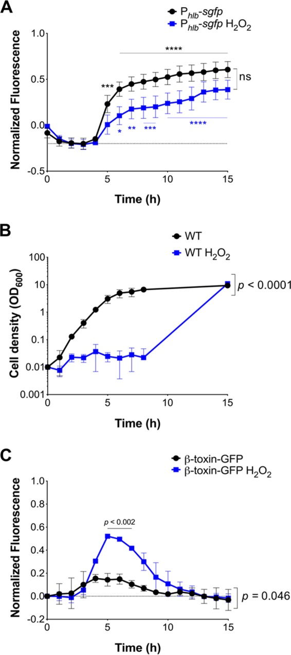
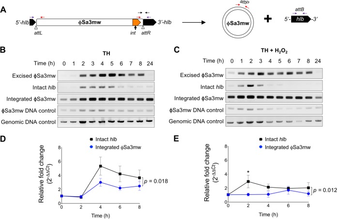
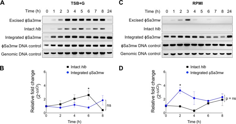
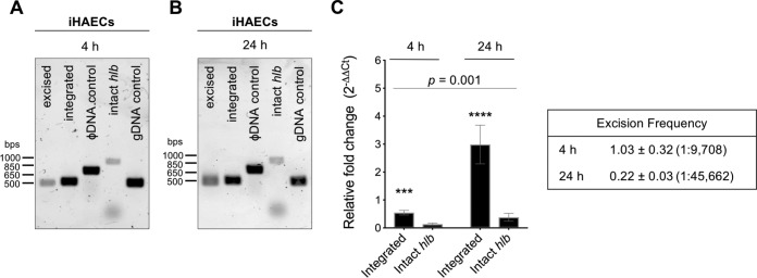
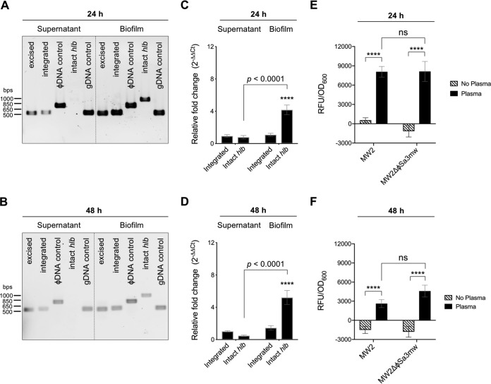
Phage regulatory switches (phage-RSs) are a newly described form of active lysogeny where prophages function as regulatory mechanisms for expression of chromosomal bacterial genes. In Staphylococcus aureus, ϕSa3int is a widely distributed family of prophages that integrate into the β-toxin structural gene hlb, effectively inactivating it. However, β-toxin-producing strains often arise during infections and are more virulent in experimental infective endocarditis and pneumonia infections. We present evidence that in S. aureus MW2, ϕSa3mw excision is temporally and differentially responsive to growth conditions relevant to S. aureus pathogenesis. PCR analyses of ϕSa3mw (integrated and excised) and of intact hlb showed that ϕSa3mw preferentially excises in response to hydrogen peroxide-induced oxidative stress and during biofilm growth. ϕSa3mw remains as a prophage when in contact with human aortic endothelial cells in culture. A criterion for a prophage to be considered a phage-RS is the inability to lyse host cells. MW2 grown under phage-inducing conditions did not release infectious phage particles by plaque assay or transmission electron microscopy, indicating that ϕSa3mw does not carry out a productive lytic cycle. These studies highlight a dynamic, and perhaps more sophisticated, S. aureus-prophage interaction where ϕSa3int prophages provide a novel regulatory mechanism for the conditional expression of virulence factors.IMPORTANCE β-Toxin is a sphingomyelinase hemolysin that significantly contributes to Staphylococcus aureus pathogenesis. In most S. aureus isolates the prophage ϕSa3int inserts into the β-toxin gene hlb, inactivating it, but human and experimental infections give rise to β-toxin-producing variants. However, it remained to be established whether ϕSa3mw excises in response to specific environmental cues, restoring the β-toxin gene sequence. This is not only of fundamental interest but also critical when designing intervention strategies and therapeutics. We provide evidence that ϕSa3mw actively excises, allowing the conditional expression of β-toxin. ϕSa3int prophages may play a novel and largely uncharacterized role in S. aureus pathogenesis as molecular regulatory switches that promote bacterial fitness and adaptation to the challenges presented by the mammalian host.

Keywords: PR-switch; Sa3int; Staphylococcus aureus; beta toxin; biofilm; infective endocarditis; oxidative stress; phage regulatory switch.


Figures

FIG 1
FIG 1
H2O2 induces β-toxin production under bacteriostatic conditions in a time-dependent manner. S. aureus wild-type (WT) and fluorescent reporter strains were grown in Todd-Hewitt (TH) medium (black) or TH plus 1 mM H2O2 (blue) overnight. (A) Fluorescence signal over time of S. aureus MW2 ectopically expressing sGFP under the control of the hlb promoter (Phlb-sgfp). (B) Bacterial cell density. (C) Fluorescence signal over time of S. aureus MW2 chromosomally expressing β-toxin fused to sGFP (β-toxin–sGFP). For panels A and C, empty vector control was subtracted from fluorescence readings at corresponding time points to correct for autofluorescence. Results are averages from at least three independent experiments performed in triplicate (means ± standard errors of the means [SEM]). Statistical significance was determined by 2-way ANOVA with Sidak’s multiple-comparison test. (A) TH, P = 0.0002 (5 h) (***) and P < 0.0001 (6 to 15 h) (****); TH plus H2O2, P = 0.023 (6 h) (*), P = 0.001 (7 h) (***), P < 0.0009 (8 to 9 h) (***), and P < 0.0001 (10 to 15 h) (****); TH versus TH plus H2O2 trend lines, not significant (ns); TH and TH plus H2O2 time effect, P < 0.0001. (B) H2O2 effect on cell density, P < 0.0001; time effect, P < 0.0001. (C) TH versus TH plus H2O2 trend lines, P = 0.046; TH versus TH plus H2O2, P < 0.0002 (5 to 7 h); TH and TH plus H2O2 time effect, P < 0.0001.
FIG 2
FIG 2
ϕSa3mw differentially excises in the presence of H2O2. (A) Schematic of ϕSa3mw integrated or excised from hlb with primer locations (colored arrows). attL and attR are the 5′- and 3′-end integration sites, respectively (arrowheads). Primer sets include excised ϕSa3mw, PCR across the attP site, present only in excised prophage DNA (red arrows); intact hlb, PCR across the attB site, present only in the absence of integrated prophage (purple arrows); integrated ϕSa3mw, PCR across the attR site, present at the 3′ end of the integration site (black arrows); ϕSa3mw DNA control, PCR within the ϕSa3mw integrase gene int (orange block arrow). (B and C) Time course PCR analysis of prophage excision in MW2 grown with aeration over a 24-h time period in Todd-Hewitt (TH) medium (B) or TH plus 1 mM H2O2 (C). Genomic DNA control was PCR within the alpha-toxin gene hla. (D and E) Time course qPCR analysis of intact hlb and integrated ϕSa3mw grown with aeration at designated time points in the presence (E) or absence (D) of 1 mM H2O2 and expressed as relative fold change from time zero. Results are averages from at least three independent experiments performed in triplicate (means ± SEM). Statistical significance was determined by 2-way ANOVA with Sidak’s multiple-comparison test. (D) Intact hlb versus integrated ϕSa3mw trend lines, P = 0.018; intact hlb and integrated ϕSa3mw time effect, P < 0.0001. (E) Intact hlb versus integrated ϕSa3mw trend lines, P = 0.012; intact hlb and integrated ϕSa3mw time effect, not significant; intact hlb versus integrated ϕSa3mw at T2h, P = 0.049 (*).
FIG 3
FIG 3
Detection of excised ϕSa3mw is greatly reduced in bacteria grown in RPMI tissue culture medium. (A and C) PCR analysis of prophage excision during MW2 planktonic growth over a 24-h time period in tryptic soy broth (TSB) supplemented with 2% glucose and 2% NaCl (TSB+G) (A) or tissue culture medium RPMI 1640 buffered with 10 mM HEPES (C). Excised ϕSa3mw, PCR across the attP site, present only in excised prophage DNA; intact hlb, PCR across the attB site, present only in the absence of integrated prophage; integrated ϕSa3mw, PCR across the attR site, present at the 3′ end of the integration site; ϕSa3mw DNA control, PCR within the ϕSa3mw integrase gene int; genomic DNA control, PCR within the alpha-toxin gene hla. (B and D) qPCR analysis of intact hlb and integrated ϕSa3mw at specific time points in TSB+G (B) or RPMI 1640 (D) and expressed as relative fold change from time zero. Results are averages from at least three independent experiments performed in triplicate (means ± SEM). Statistical significance was determined by 2-way ANOVA with Sidak’s multiple-comparison test. (B) Intact hlb versus integrated ϕSa3mw trend lines, not significant (ns); intact hlb and integrated ϕSa3mw time effect, not significant (ns); intact hlb versus integrated ϕSa3mw at the 6-h time point (T6h), P = 0.01 (*). (D) Intact hlb versus integrated ϕSa3mw trend lines, not significant (ns); intact hlb and integrated ϕSa3mw time effect, P = 0.005; intact hlb versus integrated ϕSa3mw at T2h, P = 0.01 (*).
FIG 4
FIG 4
ϕSa3mw excision is not promoted during infection of immortalized human aortic endothelial cells (iHAECs). iHAECs were infected at an MOI of 100 for 1 h, washed, and cultured for an additional 4 h and 24 h. (A and B) PCR analysis of prophage excision during iHAEC infection at 4 h (A) or 24 h (B). Excised, PCR across the ϕSa3mw attP site, present only in excised prophage DNA; integrated, PCR across the attR site, present at the 3′ end of the integration site; ϕSa3mw DNA control, PCR within the ϕSa3mw integrase gene int; intact hlb, PCR across the attB site, present only in the absence of integrated prophage; genomic DNA control (gDNA), PCR within the alpha-toxin gene hla. (C, left) qPCR analysis of integrated ϕSa3mw and intact hlb from iHAEC culture lysates at 4 h and 24 h and expressed as relative fold change from overnight cultures of MW2 grown in TH. (Right) Excision frequencies (×10−4) at 4 h and 24 h calculated as 2−ΔCT intact hlb/2−ΔCT integrated ϕSa3mw. Cell ratios were calculated as 1/excision frequency. Excision frequency at T0h, 1.38 × 10−4 ± 0.82 × 10−4 (1:7,246). Results are averages from at least three independent experiments performed in triplicate (means ± SEM). Statistical significance was determined by 2-way ANOVA with Sidak’s multiple-comparison test. (C) Integrated ϕSa3mw versus intact hlb, P = 0.0002 (4 h) (***) and P < 0.0001 (24 h) (****). Integrated ϕSa3mw and intact hlb time effect, P = 0.001.
FIG 5
FIG 5
ϕSa3mw excises during biofilm growth. (A and B) PCR analysis of prophage excision during growth in a stationary biofilm in tryptic soy broth supplemented with 2% glucose and 2% NaCl (TSB+G) for 24 h (A) or 48 h (B) and respective supernatants. Excised, PCR across the ϕSa3mw attP site, present only in excised prophage DNA; integrated, PCR across the attR site, present at the 3′ end of the integration site; ϕSa3mw DNA control, PCR within the ϕSa3mw integrase gene int; intact hlb, PCR across the attB site, present only in the absence of integrated prophage; genomic DNA control (gDNA), PCR within the alpha-toxin gene hla. (C and D) qPCR analysis of integrated ϕSa3mw and intact hlb from bacterial biofilms and supernatants at 24 h (C) and 48 h (D) and expressed as relative fold change from overnight cultures of MW2 grown in TSB+G. (E and F) Detection of β-toxin–sGFP from MW2 or MW2ΔϕSa3mw that was grown in biofilms (black bars) or planktonic (patterned bars) after 24 h (E) or 48 h (F). Results are averages from at least three independent experiments performed in triplicate (means ± SEM). Statistical significance was determined by 2-way ANOVA with Sidak’s multiple-comparison test. (C and D) Integrated ϕSa3mw in supernatants versus in biofilms at both 24 h and 48 h, not significant; intact hlb in supernatants versus in biofilms at both 24 h and 48 h, P < 0.0001; integrated ϕSa3mw versus intact hlb in biofilms at both 24 h and 48 h, P < 0.0001 (****). (E and F) MW2 RFU/OD600 in biofilms versus planktonic at both 24 h and 48 h, P < 0.0001 (****); MW2ΔϕSa3mw RFU/OD600 in biofilms versus planktonic at both 24 h and 48 h, P < 0.0001 (****); MW2 versus MW2ΔϕSa3mw RFU/OD600 in biofilms, not significant; MW2 versus MW2ΔϕSa3mw RFU/OD600 planktonic, not significant.

Similar articles

Cited by

References

    1. Salgado-Pabón W, Schlievert PM. 2014. Models matter: the search for an effective Staphylococcus aureus vaccine. Nat Rev Microbiol 12:585–591. doi:10.1038/nrmicro3308. - DOI - PubMed
    1. Wisplinghoff H, Bischoff T, Tallent SM, Seifert H, Wenzel RP, Edmond MB. 2004. Nosocomial bloodstream infections in US hospitals: analysis of 24,179 cases from a prospective nationwide surveillance study. Clin Infect Dis 39:309–317. doi:10.1086/421946. - DOI - PubMed
    1. Klein E, Smith DL, Laxminarayan R. 2007. Hospitalizations and deaths caused by methicillin-resistant Staphylococcus aureus, United States, 1999–2005. Emerg Infect Dis 13:1840–1846. doi:10.3201/eid1312.070629. - DOI - PMC - PubMed
    1. King JM, Kulhankova K, Stach CS, Vu BG, Salgado-Pabón W. 2016. Phenotypes and virulence among Staphylococcus aureus USA100, USA200, USA300, USA400, and USA600 clonal lineages. mSphere 1:e00071-16. - PMC - PubMed
    1. Alibayov B, Baba-Moussa L, Sina H, Zdeňková K, Demnerová K. 2014. Staphylococcus aureus mobile genetic elements. Mol Biol Rep 41:5005–5018. doi:10.1007/s11033-014-3367-3. - DOI - PubMed

Publication types

MeSH terms

Substances