. 2019 Apr 23;116(17):8535-8543.
doi: 10.1073/pnas.1815745116. Epub 2019 Apr 8.

Ebolavirus polymerase uses an unconventional genome replication mechanism

Affiliations

Ebolavirus polymerase uses an unconventional genome replication mechanism

Laure R Deflubé et al. Proc Natl Acad Sci U S A. .

Abstract

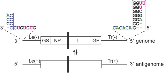
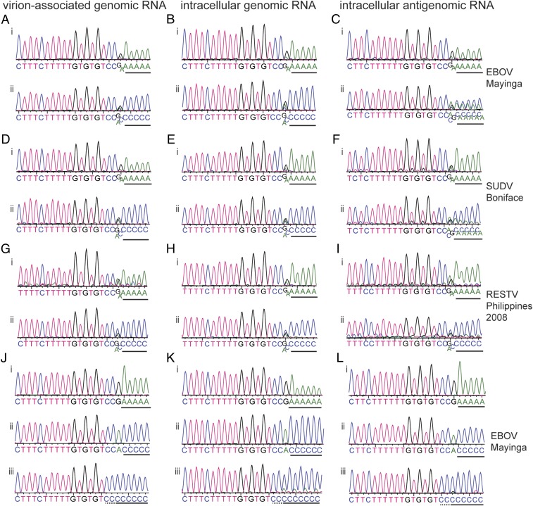
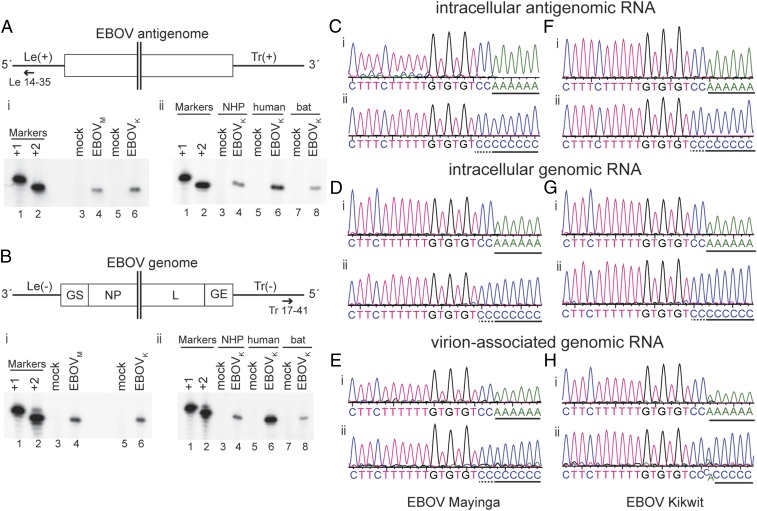
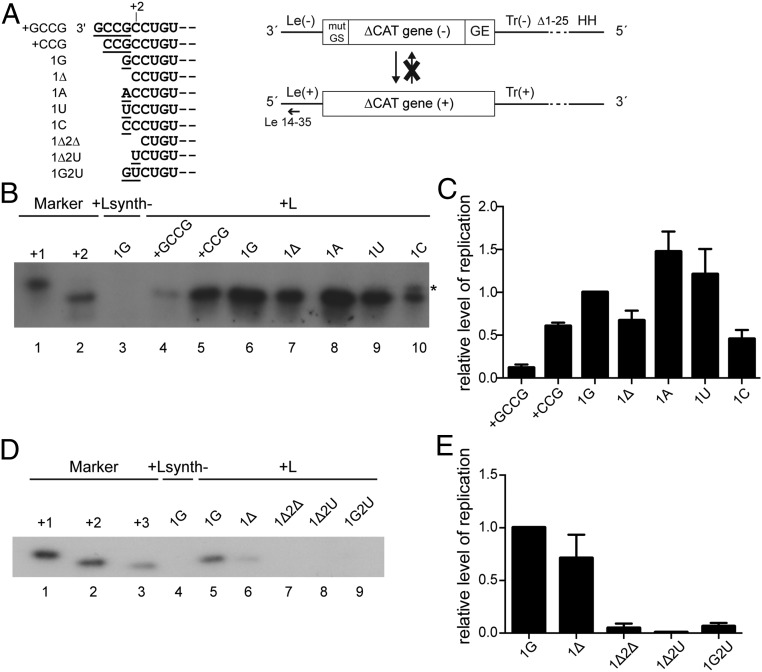
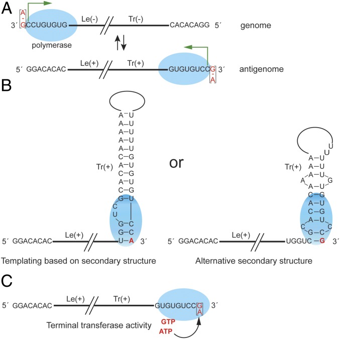
Most nonsegmented negative strand (NNS) RNA virus genomes have complementary 3' and 5' terminal nucleotides because the promoters at the 3' ends of the genomes and antigenomes are almost identical to each other. However, according to published sequences, both ends of ebolavirus genomes show a high degree of variability, and the 3' and 5' terminal nucleotides are not complementary. If correct, this would distinguish the ebolaviruses from other NNS RNA viruses. Therefore, we investigated the terminal genomic and antigenomic nucleotides of three different ebolavirus species, Ebola (EBOV), Sudan, and Reston viruses. Whereas the 5' ends of ebolavirus RNAs are highly conserved with the sequence ACAGG-5', the 3' termini are variable and are typically 3'-GCCUGU, ACCUGU, or CCUGU. A small fraction of analyzed RNAs had extended 3' ends. The majority of 3' terminal sequences are consistent with a mechanism of nucleotide addition by hairpin formation and back-priming. Using single-round replicating EBOV minigenomes, we investigated the effect of the 3' terminal nucleotide on viral replication and found that the EBOV polymerase initiates replication opposite the 3'-CCUGU motif regardless of the identity of the 3' terminal nucleotide(s) and of the position of this motif relative to the 3' end. Deletion or mutation of the first residue of the 3'-CCUGU motif completely abolished replication initiation, suggesting a crucial role of this nucleotide in directing initiation. Together, our data show that ebolaviruses have evolved a unique replication strategy among NNS RNA viruses resulting in 3' overhangs. This could be a mechanism to avoid antiviral recognition.

Keywords: Ebola virus genome ends; Ebola virus replication; ebolavirus replication initiation; nonsegmented negative strand RNA virus replication; variable 3′ genome ends.


Conflict of interest statement

The authors declare no conflict of interest.

Figures

Fig. 1.
Fig. 1.
Schematic diagram illustrating the variety of published ebolavirus 3′ and 5′ genome ends. The Le(−) region is followed by the gene start signal (GS) for the first gene, nucleoprotein (NP) gene. The Tr(−) region is preceded by the gene end signal (GE) for the L gene. Most of the viral genes are not shown. − symbols indicate the lack of a nucleotide. The complementary antigenome is depicted beneath the genome.
Fig. 2.
Fig. 2.
Sequence analysis of the 3′ ends of ebolavirus RNAs. (AI) Vero cells were infected with the indicated ebolavirus species and total cellular or virion-associated RNA was used for 3′ RACE and sequence analysis. The traces show sequences of the 3′ RACE PCR population obtained from virion-associated genomic RNA (A, D, and G) intracellular viral genomic RNA (B, E, and H), or intracellular viral antigenomic RNA (C, F, and I). In each case, i and ii show results from RNA tailed with ATP or CTP, respectively. (JL) Representative traces of the most frequently observed sequences obtained from analysis of single cDNA clones of virion-associated (J), intracellular genomic viral RNA (K), or intracellular antigenomic viral RNA (L) isolated from Vero cells infected with EBOV Mayinga. Poly(A) (i) and poly(C) (ii and iii) sequences added during the 3′ RACE procedure are underlined with a black line. It should be noted that the polymeric sequence may include some virus-specific sequence that cannot be distinguished from the poly(A) or poly(C) tail (underlined with a dotted line).
Fig. 3.
Fig. 3.
Mapping the 5′ end of EBOV RNAs by primer extension and sequence analysis. (A and B) Primer extension analysis of antigenome (A) and genome (B) RNAs. (A and B, Upper) Schematic diagram (not to scale) of the EBOV RNA that was analyzed showing the hybridization positions of the negative sense Le 14–35 (A) and positive sense Tr 17–41 (B) primers used for primer extension analysis. (A and B, Lower) Primer extension analysis. In each case, i shows analysis of RNA isolated from Vero cells infected with EBOV Mayinga (EBOVM) or Kikwit (EBOVK), and ii shows analysis of RNA isolated from EBOV Kikwit-infected Vero (nonhuman primate, NHP), Huh7 (human), and R05T (bat) cells. In each panel, [γ-32P]ATP end-labeled DNA oligonucleotides corresponding in length and sequence to cDNA representing initiation from positions +1 and +2, relative to the published EBOV sequences, were used as markers (lanes 1 and 2). (CH) Vero cells were infected with EBOV Mayinga (CE) or Kikwit (FH) and total cellular or virion-associated RNA was used for RACE analysis. The traces show sequences of the 5′ RACE PCR population obtained from the 5′ ends of intracellular antigenomic viral RNA (C and F), intracellular genomic viral RNA (D and G), or virion-associated genomic RNA (E and H). The cDNA was tailed with dATP (i) or dCTP (ii). The black line below the sequence traces indicates poly(A) or poly(C) tail sequences that had been added to the virus-specific sequence during RACE. This may include some virus-specific sequence that cannot be distinguished from the poly(A) or poly(C) tail. The first two C residues that belong to the viral sequence are underlined with a dotted line in the poly(C)-tailed sequence traces.
Fig. 4.
Fig. 4.
Analysis of replicated RNA produced from EBOV minigenomes with different 3′ ends. (A) Schematic diagram (not to scale) showing the different 3′ terminal sequences that were tested (Left) and the organization of the single-cycle replicating, nontranscribing EBOV minigenome and the antiminigenome product (Right). The terminal 25 nucleotides of the trailer were deleted (Δ1–25). The trailer is flanked by an inactive hammerhead ribozyme (HH). The annealing position of the negative sense primer used for primer extension is indicated; mut GS, mutated gene start signal; GE, gene end signal. (B) Primer extension analysis of antiminigenome RNA from minigenomes with +GCCG, +CCG, 1G, 1Δ, 1A, 1U, and 1C 3′ ends using a negative sense primer that annealed within the Le(+) region. The markers are [γ-32P]ATP end-labeled DNA oligonucleotides corresponding in length and sequence to cDNA representing initiation from positions +1 and +2, relative to published sequence. As a negative control, L was replaced with the enzymatically inactive Lsynth- mutant (52). A representative result of three independent experiments is shown. (C) Quantification of primer extension products shown in B. The data are normalized to the level of product generated by the 1G minigenome after subtraction of the Lsynth- negative control. Shown are the mean and SE of three independent experiments. (D) Primer extension analysis of antiminigenome RNA from minigenomes with 1G, 1Δ, 1Δ2Δ, 1Δ2U, and 1G2U 3′ ends using a negative sense primer that annealed within the Le(+) region. The markers are [γ-32P]ATP end-labeled DNA oligonucleotides corresponding in length and sequence to cDNA representing initiation from positions +1, +2, and +3 relative to published sequence. As a negative control, L was replaced with the enzymatically inactive Lsynth- mutant (52). A representative result of three independent experiments is shown. (E) Quantification of primer extension products shown in D. The data are normalized to the level of product generated by the 1G minigenome after subtraction of the Lsynth- negative control. Shown are the mean and SE of three independent experiments.
Fig. 5.
Fig. 5.
Model for RNA synthesis initiation and 3′ terminal nucleotide addition by the ebolavirus polymerase. (A) Scheme showing the site of initiation at the Le(−) promoter of the genome and at the Tr(+) promoter of the antigenome. The ebolavirus polymerase is indicated by a blue ellipse and the initiation site by a green arrow. The experimentally determined 3′ terminal nucleotide is highlighted in red. − symbols indicate the lack of a nucleotide. (B) Schematic diagram showing how addition of a G or A residue onto the 3′ end of antigenome RNA may occur by templating of the 3′ terminal nucleotide using secondary structure formations at the 3′ end of the RNA. (C) Schematic diagram showing addition of a 3′ terminal purine nucleotide by terminal nucleotidyl transferase activity of the ebolavirus polymerase or a cellular terminal transferase.

Comment in

  • Ebola Virus Replication Stands Out.
    Wang W, Wu C, Amarasinghe GK, Leung DW. Wang W, et al. Trends Microbiol. 2019 Jul;27(7):565-566. doi: 10.1016/j.tim.2019.05.004. Epub 2019 May 30. Trends Microbiol. 2019. PMID: 31155428 Free PMC article.

Similar articles

Cited by

References

    1. Whelan SP, Barr JN, Wertz GW. Transcription and replication of nonsegmented negative-strand RNA viruses. Curr Top Microbiol Immunol. 2004;283:61–119. - PubMed
    1. Kiley MP, Wilusz J, McCormick JB, Keene JD. Conservation of the 3′ terminal nucleotide sequences of Ebola and Marburg virus. Virology. 1986;149:251–254. - PubMed
    1. Volchkov VE, et al. Characterization of the L gene and 5′ trailer region of Ebola virus. J Gen Virol. 1999;80:355–362. - PubMed
    1. Weik M, Enterlein S, Schlenz K, Mühlberger E. The Ebola virus genomic replication promoter is bipartite and follows the rule of six. J Virol. 2005;79:10660–10671. - PMC - PubMed
    1. Kolesnikova L, Nanbo A, Becker S, Kawaoka Y. Inside the cell: Assembly of filoviruses. Curr Top Microbiol Immunol. 2017;411:353–380. - PubMed

Publication types