. 2019 Apr 8;9(1):5770.
doi: 10.1038/s41598-019-42203-z.

Moderate exercise improves function and increases adiponectin in the mdx mouse model of muscular dystrophy

Affiliations

Moderate exercise improves function and increases adiponectin in the mdx mouse model of muscular dystrophy

Aaron S Zelikovich et al. Sci Rep. .

Abstract

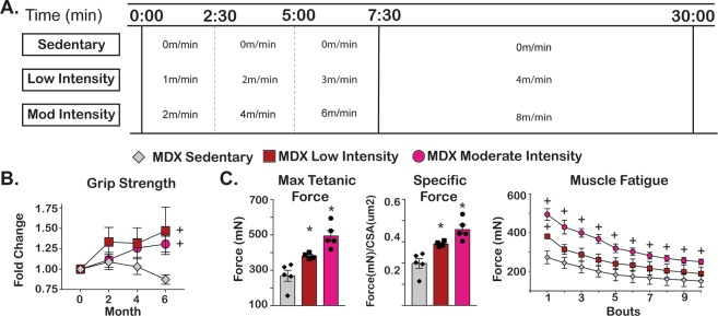
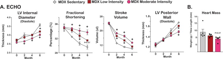
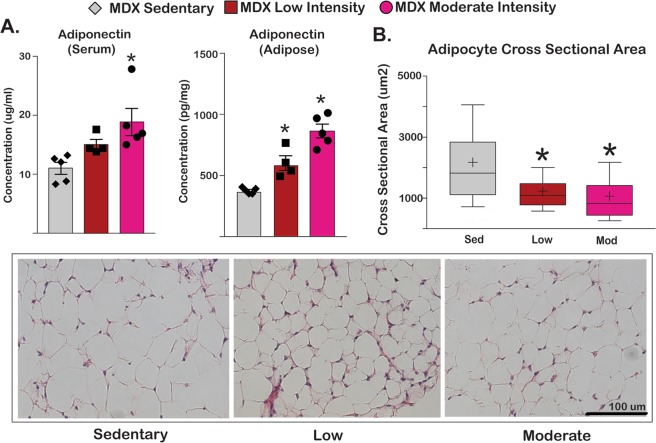
The loss of dystrophin produces a mechanically fragile sarcolemma, causing muscle membrane disruption and muscle loss. The degree to which exercise alters muscular dystrophy has been evaluated in humans with Duchenne Muscular Dystrophy (DMD) and in mouse models including the mdx mouse but with inconsistent findings. We now examined two different levels of exercise, moderate and low intensity, in the mdx mouse model in the DBA2J background. mdx mice at 4-5 months of age were subjected to two different doses of exercise. We found a dose-dependent benefit for low and moderate exercise, defined as 4 m/min or 8 m/min, for 30 minutes three times a week. After six months, exercised mdx mice showed improved tetanic and specific force compared to the sedentary group. We also observed increased respiratory capacity manifesting as greater minute volume, as well as enhanced cardiac function mitigating the decline of fractional shortening that is normally seen. Exercised mdx mice also showed a dose-dependent increase in serum adiponectin with a concomitant reduced adipocyte cross sectional area. These findings identify moderate intensity exercise as a means to improve muscle performance in the mdx DBA2J mice and suggest serum adiponectin as a biomarker for beneficial exercise effect in DMD.


Conflict of interest statement

The authors declare no competing interests.

Figures

Figure 1
Figure 1
Exercise improved skeletal muscle function in mdx mice. Four month-old male mdx mice from the DBA2J background (n = 5/cohort) were exercised three times per week for 24 weeks between 8 am–12 pm. (A) The exercise protocol included a 7 min 30 second warm-up with an increase in speed every 2 minutes and 30 seconds until maximum speed was attained. Mice remained on treadmill for additional 22 minutes and 30 seconds at maximum speed for a total of 30 minutes duration. Three levels of exercise were evaluated: 0 m/min (sedentary), 4 m/min (low intensity) and 8 m/min (moderate intensity). (B) Average forelimb grip strength increased in both exercise groups whereas it declined in sedentary group. (C) Maximum tetanic force and specific force was determined for the Tibialis anterior (TA) muscles and showed an intensity-dependent improvement in muscle function that was sustained over 10 consecutive bouts of isometric contraction. Histograms depict single values and mean ± s.e.m.; curves depict mean ± s.e.m.; box plots depict 10–90% distribution with mean(+); *P < 0.05 vs sedentary; 1-way ANOVA test with Tukey’s multiple comparison; + , P < 0.05 vs vehicle, 2-way ANOVA test with Tukey’s multiple comparison.
Figure 2
Figure 2
Exercise improved respiratory function in mdx mice. Whole-body plethysmography demonstrated an increase in respiratory minute volume in an intensity-dependent manner. Inspiratory (Ti) and expiratory (Te) times were inversely related to disease progression. Both exercise groups had an attenuation of increased Ti and Te as compared to sedentary group. Curves depict mean ± s.e.m.; + , P < 0.05 vs vehicle, 2-way ANOVA test with Tukey’s multiple comparison.
Figure 3
Figure 3
Exercise attenuated cardiac decline associated with disease progression in mdx mice. (A) Echocardiographic parameters indicated attenuation of cardiac decline associated with disease progression in exercised groups. Reduced left ventricular diameter, improved fractional shortening, and increased stroke volume were present after exercise. Hypertrophy associated with disease progression was attenuated with a smaller posterior wall thickness and (B) Heart mass showed a modest reduction in exercised mdx mice. Histograms depict single values and mean ± s.e.m.; curves depict mean ± s.d.; curves depict mean ± s.e.m.; *P < 0.05 vs sedentary; 1-way ANOVA test with Tukey’s multiple comparison; + , P < 0.05 vs sedentary, 2-way ANOVA test with Tukey’s multiple comparison.
Figure 4
Figure 4
Exercise did not increase fibrosis in mdx mice. (A–C) Heart, diaphragm, and gastrocnemius showed no increase in Sirius red staining with exercise. Hydroxyproline content did not differ among groups confirming the histological findings.
Figure 5
Figure 5
Exercise increased serum adiponectin and decreased adipocyte cross sectional area in mdx mice. (A) Serum adiponectin levels and adiponectin content in gastrocnemius (GCN) muscle and adipose tissue were increased in the exercise cohorts. (B) Adipocyte cross sectional area decreased in exercised mdx mice, as quantitated from histological staining of omental fat. Histograms depict single values and mean ± s.e.m.; box plots depict 10–90% distribution with mean (+); *P < 0.05 vs sedentary; 1-way ANOVA test with Tukey’s multiple comparison.
Figure 6
Figure 6
Moderate intensity treadmill running was associated with increased activity in mdx mice (A) An Activity Index (AI) was used to compare activity before and after treadmill exercise. A score of 1 was given per each time a mouse crossed the treadmill midline, and the score pre-exercise was subtracted from the post-exercise score. (B,C) Sustained activity post exercise was only seen in moderate intensity exercise group. (D) mdx mice from the DBA2J background were subjected to low or moderate intensity exercise over four days, and this exposure to exercise did not alter activity. (E) Strain-matched WT and mdx-DBA2J mice were exercised over four days using the low intensity protocol. Comparing activity levels after exercise from day 4 to day 0 showed activity was greater in WT mice versus mdx mice. Activity was higher in WT than in mdx mice when monitored for 5-minutes post-exercise at day 4. Histograms depict single values and mean ± s.e.m.; curves depict mean ± s.e.m.; *P < 0.05 vs sedentary; 1-way ANOVA test with Tukey’s multiple comparison; ns (non-significant), P > 0.05, + , P < 0.05 vs sedentary, 2-way ANOVA test with Tukey’s multiple comparison.

References

    1. Guiraud S, et al. The Pathogenesis and Therapy of Muscular Dystrophies. Annu Rev Genomics Hum Genet. 2015;16:281–308. doi: 10.1146/annurev-genom-090314-025003. - DOI - PubMed
    1. Eagle M. Report on the muscular dystrophy campaign workshop: exercise in neuromuscular diseases Newcastle, January 2002. Neuromuscul Disord. 2002;12:975–983. doi: 10.1016/S0960-8966(02)00136-0. - DOI - PubMed
    1. Wineinger MA, Abresch RT, Walsh SA, Carter GT. Effects of aging and voluntary exercise on the function of dystrophic muscle from mdx mice. American journal of physical medicine & rehabilitation. 1998;77:20–27. doi: 10.1097/00002060-199801000-00004. - DOI - PubMed
    1. Sandri M, et al. Exercise induces myonuclear ubiquitination and apoptosis in dystrophin-deficient muscle of mice. Journal of neuropathology and experimental neurology. 1997;56:45–57. doi: 10.1097/00005072-199701000-00005. - DOI - PubMed
    1. Hulmi JJ, et al. Exercise restores decreased physical activity levels and increases markers of autophagy and oxidative capacity in myostatin/activin-blocked mdx mice. American journal of physiology. Endocrinology and metabolism. 2013;305:E171–182. doi: 10.1152/ajpendo.00065.2013. - DOI - PubMed

Publication types

LinkOut - more resources