. 2019 May;22(5):820-827.
doi: 10.1038/s41593-019-0371-x. Epub 2019 Apr 8.

Working memory revived in older adults by synchronizing rhythmic brain circuits

Affiliations

Working memory revived in older adults by synchronizing rhythmic brain circuits

Robert M G Reinhart et al. Nat Neurosci. 2019 May.

Abstract

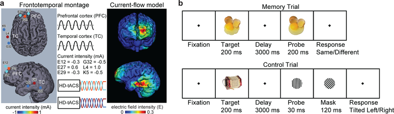
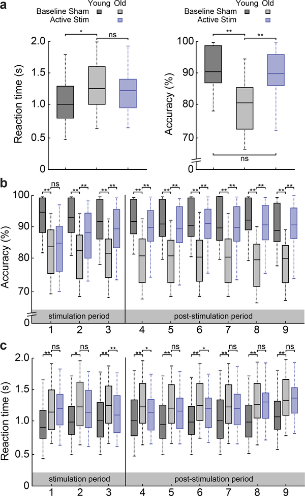
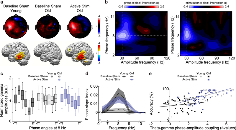
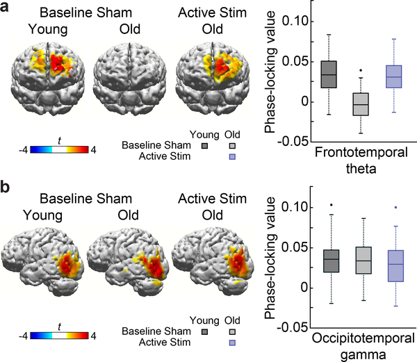
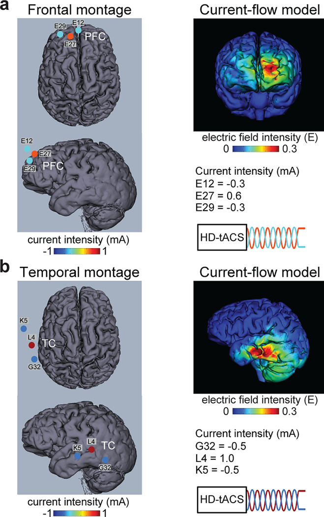
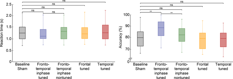
Understanding normal brain aging and developing methods to maintain or improve cognition in older adults are major goals of fundamental and translational neuroscience. Here we show a core feature of cognitive decline-working-memory deficits-emerges from disconnected local and long-range circuits instantiated by theta-gamma phase-amplitude coupling in temporal cortex and theta phase synchronization across frontotemporal cortex. We developed a noninvasive stimulation procedure for modulating long-range theta interactions in adults aged 60-76 years. After 25 min of stimulation, frequency-tuned to individual brain network dynamics, we observed a preferential increase in neural synchronization patterns and the return of sender-receiver relationships of information flow within and between frontotemporal regions. The end result was rapid improvement in working-memory performance that outlasted a 50 min post-stimulation period. The results provide insight into the physiological foundations of age-related cognitive impairment and contribute to groundwork for future non-pharmacological interventions targeting aspects of cognitive decline.


Conflict of interest statement

Competing interests

The authors declare no competing interests.

Figures

Fig. 1:
Fig. 1:. Experiment 1, frontotemporal HD-tACS procedure and task.
a, The multifocal inphase frontotemporal HD-tACS montage and current-flow model shown on 3D reconstructions of the cortical surface. The location and current intensity value of each stimulating electrode are shown. The left prefrontal cortex and left temporal cortex were targeted, each with three electrodes in a center-surround, source-sink pattern to achieve maximum focality. b, All trials began with fixation followed by a target and then a delay period. In memory blocks, subjects judged whether a later probe was the same or a modified version of the target. In control blocks, subjects judged whether a backward masked grating probe was tilted clockwise or counterclockwise from the vertical and were not required to remember the target. Trial-by-trial adjustments in orientation magnitude of the probe ensured comparable performance across memory and control blocks.
Fig. 2:
Fig. 2:. Experiment 1, behavioral results.
a, Box plots of reaction time (RT) from correct trials and accuracy in post-stimulation memory blocks show older adults were slower (t82 = 2.331, p = 0.022, dz = 0.509) and less accurate (t82 = 5.587, p < 0.01, dz = 1.219) at baseline, relative to younger adults. After stimulation, accuracy improved (t41 = 3.738, p = 0.001, dz = 0.577), removing the difference between groups (t82 = 0.939, p = 0.350, dz = 0.205). RT did not differ between conditions (t41 = 0.641, p = 0.527, dz = 0.099). Box plots of accuracy (b) and RT (c) from memory blocks sorted into 9 sequential bins (4 minutes per bin with 4 minutes between each bin from interleaved control blocks) shows significant group differences at every time bin for accuracy (ts82 > 6.824, ps < 0.01, dsz > 1.489) and RT (ts82 > 2.432, ps < 0.018, dsz > 0.531), at baseline. Stimulation improved accuracy at every time bin (ts41 > 4.393, ps < 0.01, dsz > 0.678), relative to sham, except the first (t41 = 0.986, p = 0.330, dz = 0.152). Stimulation briefly sped RT at time bins 3 (t41 = 2.841, p = 0.007, dz = 0.438), 4 (t41 = 2.278, p = 0.028, dz = 0.352), and 6 (t41 = 2.282, p = 0.028, dz = 0.352). Between-group comparisons used independent samples two-tailed t-tests (n = 84). Within-group comparisons used paired sample two-tailed t-tests (n = 42). Box-plot center, median; box limits, lower and upper quartiles; whiskers, lower and upper extreme values. * p < 0.05. ** p < 0.01.
Fig. 3:
Fig. 3:. Experiment 1, phase-amplitude coupling results.
a, Results show a significantly increased phase-amplitude coupling (PAC) cluster in left temporal electrodes (top) and voxels (bottom) for theta (7–9 Hz) phase and gamma (26–34 Hz) amplitude frequencies on memory compared to control blocks in younger adults at baseline and older adults after frontotemporal inphase theta HD-tACS (pcorrected < 0.01, two-sided cluster permutation test, n = 42). No other clusters were identified. b, Comodulograms show group x block and stimulation x block interactions for all combinations of low-frequency phase (2–16 Hz) and high-frequency amplitude (18–120 Hz) from left temporal electrodes showing significant PAC. Crosshairs indicate values where t > −2.6 or −2.4, respectively (pcorrected < 0.01, two-sided permutation test, n = 42). c, Box plots of gamma (26–34 Hz) amplitude binned by 8-Hz phase shown across groups and conditions (n = 42). d, Cross-frequency directionality between 6.5–10.5-Hz phase and 26–34-Hz amplitude from left temporal electrodes for each group and condition (center, mean; shaded region, SEM; n = 42). e, Individual subject Pearson correlations (two-tailed) between accuracy and theta-gamma PAC from left temporal electrodes for each group and condition (n = 42). Box-plot center, median; box limits, lower and upper quartiles; whiskers, lower and upper extreme values; points, outliers.
Fig. 4:
Fig. 4:. Experiment 1, phase synchronization results.
a, A significant cluster in prefrontal cortex showed increased phase-locked value (PLV) with temporal cortex seed region in the theta band for younger adults at baseline and for older adults after stimulation (pcorrected < 0.01, two-sided cluster permutation test, n = 42). For theta PLV, the between-group difference at baseline (t82 = 6.450, p < 0.01, dz = 0.995) and the between-condition difference within older adults (t41 = 8.238, p < 0.01, dz = 1.271) were significant. b, A significant cluster in lateral occipital cortex showed increased PLV with temporal cortex seed region in the gamma band for each group and condition (pcorrected < 0.01, two-sided cluster permutation test, n = 42). For gamma PLV, there were no significant group (t82 = 0.898, p = 0.374, dz = 0.139) or condition differences (t41 = 1.012, p = 0.317, dz = 0.156). No other clusters were found. Between-group comparisons used independent two-tailed t-tests (n = 84). Within-group comparisons used paired sample two-tailed t-tests (n = 42). Box-plot center, median; box limits, lower and upper quartiles; whiskers, lower and upper extreme values; points, outliers.
Fig. 5:
Fig. 5:. Experiment 2, single-region HD-tACS procedures.
The unifocal frontal (a) and unifocal temporal (b) HD-tACS montages and current-flow models shown on 3D reconstructions of the cortical surface. The location and current intensity value of each stimulating electrode are shown. The frontal montage targeted left prefrontal cortex. The temporal montage targeted left temporal cortex. Each stimulation site used three electrodes in a center-surround, source-sink pattern to achieve maximum focality.
Fig. 6:
Fig. 6:. Experiment 2, behavioral results.
Box plots of reaction time (RT) from correct trials and accuracy of older adults from post-stimulation memory blocks shown across the stimulation conditions of Experiment 2. Relative to sham, frontotemporal inphase theta-tuned stimulation exerted a preferential improvement in task accuracy (t27 = 3.101, p = 0.004, dz = 0.586) without changing RT (t27 = 1.278, p = 0.212, dz = 0.242). Nontuned stimulation had no significant impact on accuracy (t27 = 1.211, p = 0.236, dz = 0.229), or RT (t27 = 0.406, p = 0.688, dz = 0.077), relative to sham. Frontal-alone and temporal-alone stimulation had no significant effect on RT (frontal, t27 = 0.182, p = 0.857, dz = 0.034; temporal, t27 = 0.085, p = 0.933, dz = 0.016) or accuracy (frontal, t27 = 0.323, p = 0.749, dz = 0.061; temporal, t27 = 0.162, p = 0.873, dz = 0.031). Paired sample two-tailed t-tests (n = 28). Box-plot center, median; box limits, lower and upper quartiles; whiskers, lower and upper extreme values. ** p < 0.01.

Comment in

References

    1. Bishop NA, Lu T & Yankner BA Neural mechanisms of ageing and cognitive decline. Nature 464, 529–535 (2010). - PMC - PubMed
    1. Hebert LE, Weuve J, Scherr PA & Evans DA Alzheimer disease in the United States (2010–2050) estimated using the 2010 Census. Neurology 80, 1778–1783 (2013). - PMC - PubMed
    1. Hayutin AM Global demographic shifts create challenges and opportunities. PREA Quart, 46–53 (2007).
    1. Park DC & Reuter-Lorenz PA The adaptive brain: aging and neurocognitive scaffolding. Nature Reviews Neuroscience 60, 173–196 (2009). - PMC - PubMed
    1. Grady C Trends in neurocognitive aging. Nature Reviews Neuroscience 13, 491–505 (2012). - PMC - PubMed

Publication types