

Elevated nuclear auto-antigenic sperm protein promotes melanoma progression by inducing cell proliferation
- PMID: 30962692
- PMCID: PMC6433116
- DOI: 10.2147/OTT.S197813
Elevated nuclear auto-antigenic sperm protein promotes melanoma progression by inducing cell proliferation
Abstract
Background: Nuclear auto-antigenic sperm protein (NASP) has been implicated in tumorigenesis. However, its role in melanoma is still unclear.
Materials and methods: In the present study, we detected the mRNA and protein level of NASP in melanoma cell lines and tissues. Then the role of NASP was investigated by transfecting with NASP siRNAs. Finally, the prognosis of NASP was analyzed in 100 melanoma patients through Cox regression and Kaplan-Meier analyses.
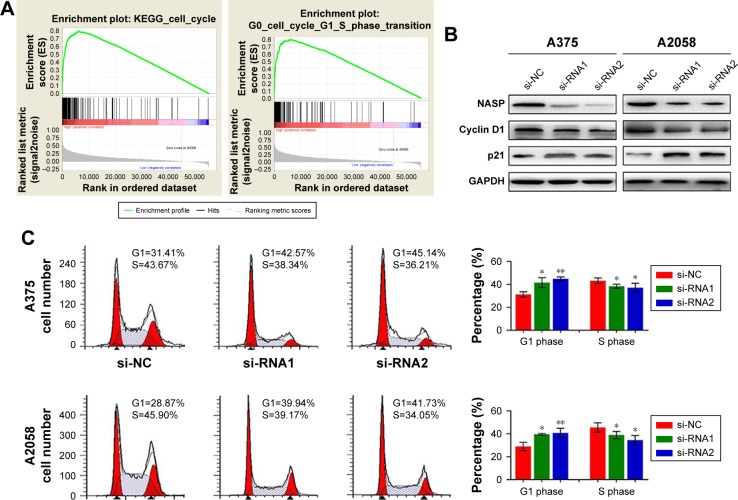
Results: We showed that NASP was significantly overexpressed in melanoma tissues, and unregulated NASP promoted melanoma cell proliferation via promoting cell cycle G1/S phase transition. Additionally, the expression of NASP was closely related to proliferating cell nuclear antigen, a widely accepted biomarker for cell proliferation. Clinically, we found that a high level of NASP predicated poor overall survival and high cumulative recurrence rates. Multivariate analysis revealed that NASP was a risk biomarker for predicting the prognosis of melanoma patients.
Conclusion: Elevated NASP plays an important role in melanoma cell proliferation and tumor progression, and it can be used as an independent prognostic biomarker for melanoma patients.
Keywords: NASP; melanoma; prognosis; proliferation.
Conflict of interest statement
Disclosure The authors report no conflicts of interest in this work.
Figures


Similar articles
-
Knockdown of long noncoding RNA colorectal neoplasia differentially expressed inhibits hepatocellular carcinoma progression by mediating the expression of nuclear autoantigenic sperm protein.Oncol Rep. 2021 Dec;46(6):252. doi: 10.3892/or.2021.8203. Epub 2021 Oct 11. Oncol Rep. 2021. PMID: 34633056 Free PMC article.
-
Somatic nuclear auto-antigenic sperm protein sensitizes human breast cancer cells to 5-Fluorouracil.Cancer Chemother Pharmacol. 2022 Apr;89(4):559-564. doi: 10.1007/s00280-021-04391-2. Epub 2022 Feb 8. Cancer Chemother Pharmacol. 2022. PMID: 35133490
-
Downregulation of tNASP inhibits proliferation through regulating cell cycle-related proteins and inactive ERK/MAPK signal pathway in renal cell carcinoma cells.Tumour Biol. 2015 Jul;36(7):5209-14. doi: 10.1007/s13277-015-3177-9. Epub 2015 Feb 12. Tumour Biol. 2015. PMID: 25669170
-
Requirement for nuclear autoantigenic sperm protein mRNA expression in bovine preimplantation development.Anim Sci J. 2016 Mar;87(3):457-61. doi: 10.1111/asj.12538. Epub 2015 Dec 22. Anim Sci J. 2016. PMID: 26690724
-
Analysis of gene expression profiles in HeLa cells in response to overexpression or siRNA-mediated depletion of NASP.Reprod Biol Endocrinol. 2009 May 13;7:45. doi: 10.1186/1477-7827-7-45. Reprod Biol Endocrinol. 2009. PMID: 19439102 Free PMC article.
Cited by
-
Nuclear Autoantigenic Sperm Protein Promotes Cardiac Regeneration and Repair Through Activating the PDGFRB/AKT Pathway.FASEB J. 2025 Aug 15;39(15):e70935. doi: 10.1096/fj.202501385R. FASEB J. 2025. PMID: 40787796 Free PMC article.
-
Nuclear autoantigenic sperm protein facilitates glioblastoma progression and radioresistance by regulating the ANXA2/STAT3 axis.CNS Neurosci Ther. 2024 Apr;30(4):e14709. doi: 10.1111/cns.14709. CNS Neurosci Ther. 2024. PMID: 38605477 Free PMC article.
-
NASP Promotes Triple-negative Breast Cancer Progression and Metastasis by Stabilizing YAP in a USP15-Dependent Way.Int J Biol Sci. 2025 Jun 20;21(9):4172-4186. doi: 10.7150/ijbs.99438. eCollection 2025. Int J Biol Sci. 2025. PMID: 40612673 Free PMC article.
References
-
- Jemal A, Siegel R, Ward E, et al. Cancer statistics, 2006. CA Cancer J Clin. 2006;56:106–130. - PubMed
LinkOut - more resources
Full Text Sources
Miscellaneous
