

Surgical management of intramuscular hemangioma of the foot: a case report
- PMID: 30962823
- PMCID: PMC6434622
- DOI: 10.1186/s13037-019-0197-1
Surgical management of intramuscular hemangioma of the foot: a case report
Abstract
Background: Hemangiomas are benign tumors usually found in the lower extremity yet their surgical management on the location in the foot is rarely documented.
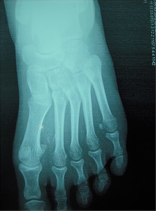
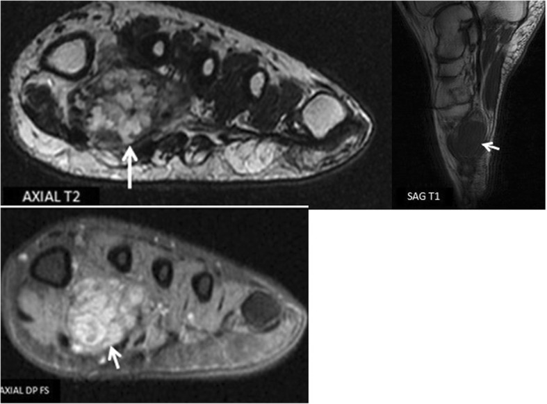
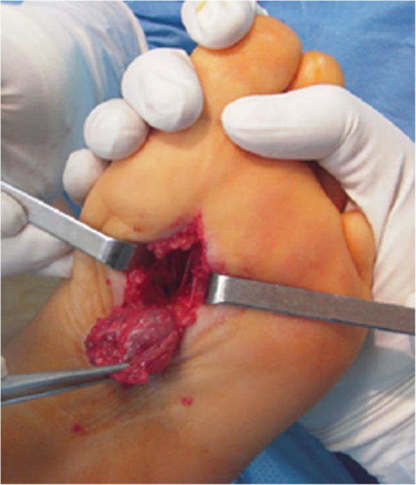
Case presentation: We report a case of a plantar intramuscular hemangioma in 25-year-old patient with a history of percutaneous therapy. Patient had undergone intralesional sclerotherapy 3 years prior to his admission with persistent pain on weight bearing activities.MRI demonstrated a multi lobulated lesion of the 1st IMS with a peripheral enhancement on gadolinium injection. The patient underwent elective surgery with complete excision and no functional impairment at the last follow-up 3 years after surgery.
Conclusion: Intramuscular hemangiomas are rare occurrences. Steroid injection and sclerotherapy are effective non-operative methods. Complete excision of isolated hemangioma lesions allows definite diagnosis with no recurrence.
Keywords: Excision; Hemangioma; Intramuscular; Plantar; Sclerotherapy.
Conflict of interest statement
The approval of the ethics committee of the Hassan II Teaching Hospital, was obtained prior to the publication of this article.Written informed consent was obtained from the patient for publication of this article and associated images.The authors declare no potential conflicts of interest with respect to the authorship, and/or publication of this article.Springer Nature remains neutral with regard to jurisdictional claims in published maps and institutional affiliations.
Figures
References
-
- Yu J, Tran D, Newhard HM. Multicompartment intramuscular hemangioma of the foot: a case study. J Am Podiatr Med Assoc 2014;104(2):203–207. 10.7547/0003-0538-104.2.203 [Pubmed] . - PubMed
-
- Mitsionis GI, Pakos EE, Kosta P, Batistatou A, Beris A. Intramuscular hemangioma of the foot: a case report and review of the literature. Foot Ankle Surg 2010;16(2):e27–e29. 10.1016/j.fas.2009.05.008. Epub 2009 Jul 7. [Pubmed]. - PubMed
-
- Davies JL, Stone PA, McGarry JJ. Mixed cavernous and capillary intraosseous hemangioma of the foot. J Am Podiatr Med Assoc 1997;87(10):478–82. [Pubmed]. - PubMed
Publication types
LinkOut - more resources
Full Text Sources
