

Application of Imaging Technologies in Breast Cancer Detection: A Review Article
- PMID: 30962849
- PMCID: PMC6447343
- DOI: 10.3889/oamjms.2019.171
Application of Imaging Technologies in Breast Cancer Detection: A Review Article
Abstract
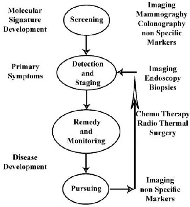
One of the techniques utilised in the management of cancer in all stages is multiple biomedical imaging. Imaging as an important part of cancer clinical protocols can provide a variety of information about morphology, structure, metabolism and functions. Application of imaging technics together with other investigative apparatus including in fluids analysis and vitro tissue would help clinical decision-making. Mixed imaging techniques can provide supplementary information used to improve staging and therapy planning. Imaging aimed to find minimally invasive therapy to make better results and reduce side effects. Probably, the most important factor in reducing mortality of certain cancers is an early diagnosis of cancer via screening based on imaging. The most common cancer in women is breast cancer. It is considered as the second major cause of cancer deaths in females, and therefore it remained as an important medical and socio-economic issue. Medical imaging has always formed part of breast cancer care and has used in all phases of cancer management from detection and staging to therapy monitoring and post-therapeutic follow-up. An essential action to be performed in the preoperative staging of breast cancer based on breast imaging. The general term of breast imaging refers to breast sonography, mammography, and magnetic resonance tomography (MRT) of the breast (magnetic resonance mammography, MRM). Further development in technology will lead to increase imaging speed to meet physiological processes requirements. One of the issues in the diagnosis of breast cancer is sensitivity limitation. To overcome this limitation, complementary imaging examinations are utilised that traditionally includes screening ultrasound, and combined mammography and ultrasound. Development in targeted imaging and therapeutic agents calls for close cooperation among academic environment and industries such as biotechnological, IT and pharmaceutical industries.
Keywords: Breast Imaging; Breast Scintimammography; Cancer detection; Mammography; Ultrasonography MRI.
Figures
Similar articles
-
Mammography screening: A major issue in medicine.Eur J Cancer. 2018 Feb;90:34-62. doi: 10.1016/j.ejca.2017.11.002. Epub 2017 Dec 20. Eur J Cancer. 2018. PMID: 29272783
-
The role of various modalities in breast imaging.Biomed Pap Med Fac Univ Palacky Olomouc Czech Repub. 2007 Dec;151(2):209-18. doi: 10.5507/bp.2007.036. Biomed Pap Med Fac Univ Palacky Olomouc Czech Repub. 2007. PMID: 18345253 Review.
-
Imaging and cancer: a review.Mol Oncol. 2008 Aug;2(2):115-52. doi: 10.1016/j.molonc.2008.04.001. Epub 2008 May 10. Mol Oncol. 2008. PMID: 19383333 Free PMC article. Review.
-
Responding to the challenges of breast cancer in egypt and other arab countries.J Egypt Natl Canc Inst. 2008 Dec;20(4):309-12. J Egypt Natl Canc Inst. 2008. PMID: 20571588
-
[New trends and novel possibilities in the diagnostic imaging of breast cancer].Magy Onkol. 2015 Mar;59(1):44-55. Epub 2014 Oct 13. Magy Onkol. 2015. PMID: 25763913 Review. Hungarian.
Cited by
-
Shear Wave Elastography in Breast Cancer: Unveiling Correlations With Histopathological Grades and Subtypes.Cureus. 2024 Jul 3;16(7):e63759. doi: 10.7759/cureus.63759. eCollection 2024 Jul. Cureus. 2024. PMID: 39099972 Free PMC article.
-
Diagnostic Accuracy of Contrast-Enhanced, Spectral Mammography (CESM) and 3T Magnetic Resonance Compared to Full-Field Digital Mammography plus Ultrasound in Breast Lesions: Results of a (Pilot) Open-Label, Single-Centre Prospective Study.Cancers (Basel). 2022 Mar 7;14(5):1351. doi: 10.3390/cancers14051351. Cancers (Basel). 2022. PMID: 35267659 Free PMC article.
-
Can Scintimammography Help Differentiate the Nature of Suspected Masses Identified in Breast Ultrasound among Young Patients?World J Nucl Med. 2025 Mar 9;24(2):155-160. doi: 10.1055/s-0045-1805044. eCollection 2025 Jun. World J Nucl Med. 2025. PMID: 40336858 Free PMC article.
-
Enhancing breast cancer diagnosis: transfer learning on DenseNet with neural hashing for histopathology fine-grained image classification.Med Biol Eng Comput. 2025 Apr 6. doi: 10.1007/s11517-025-03346-6. Online ahead of print. Med Biol Eng Comput. 2025. PMID: 40189728
-
Artificial intelligence in breast cancer: application and future perspectives.J Cancer Res Clin Oncol. 2023 Nov;149(17):16179-16190. doi: 10.1007/s00432-023-05337-2. Epub 2023 Sep 1. J Cancer Res Clin Oncol. 2023. PMID: 37656245 Free PMC article. Review.
References
-
- Siegel R, Ma J, Zou Z, et al. Cancer statistics, 2014. CA Cancer J Clin. 2014;64:9e29. - PubMed
-
- DeSantis CE, Lin CC, Mariotto AB, et al. Cancer treatment and survivorship statistics, 2014. CA Cancer J Clin. 2014;64:252e71. - PubMed
-
- Forootan M, Tabatabaeefar M, Mosaffa N, Rahimzadeh Ashkalak H, Darvishi M. Investigating Esophageal Stent-Placement Outcomes in Patients with Inoperable Non-Cervical Esophageal Cancer. J Cancer. 2018;9(1):213–218. https://doi.org/10.7150/jca.21854 PMid:29290788 PMCid:PMC5743730. - PMC - PubMed
-
- Fass L. Imaging and cancer:a review. Molecular oncology. 2008;2(2):115–52. https://doi.org/10.1016/j.molonc.2008.04.001 PMid:19383333 PMCid:PMC5527766. - PMC - PubMed
-
- Ehman RL, Hendee WR, Welch MJ, Dunnick NR, Bresolin LB, Arenson RL, Baum S, Hricak H, Thrall JH. Blueprint for imaging in biomedical research. Radiology. 2007;244(1):12–27. https://doi.org/10.1148/radiol.2441070058 PMid:17507725. - PubMed
Publication types
LinkOut - more resources
Full Text Sources