. 2019 Mar 6:2019:2039856.
doi: 10.1155/2019/2039856. eCollection 2019.

Protective Role of Nuclear Factor Erythroid-2-Related Factor 2 against Mechanical Trauma-Induced Apoptosis in a Vaginal Distension-Induced Stress Urinary Incontinence Mouse Model

Affiliations

Protective Role of Nuclear Factor Erythroid-2-Related Factor 2 against Mechanical Trauma-Induced Apoptosis in a Vaginal Distension-Induced Stress Urinary Incontinence Mouse Model

Jianming Tang et al. Oxid Med Cell Longev. .

Abstract

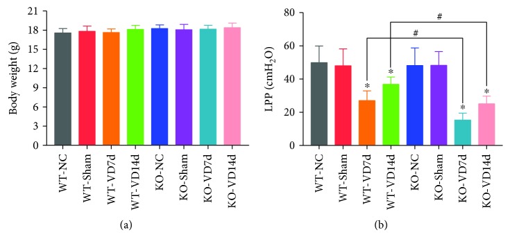
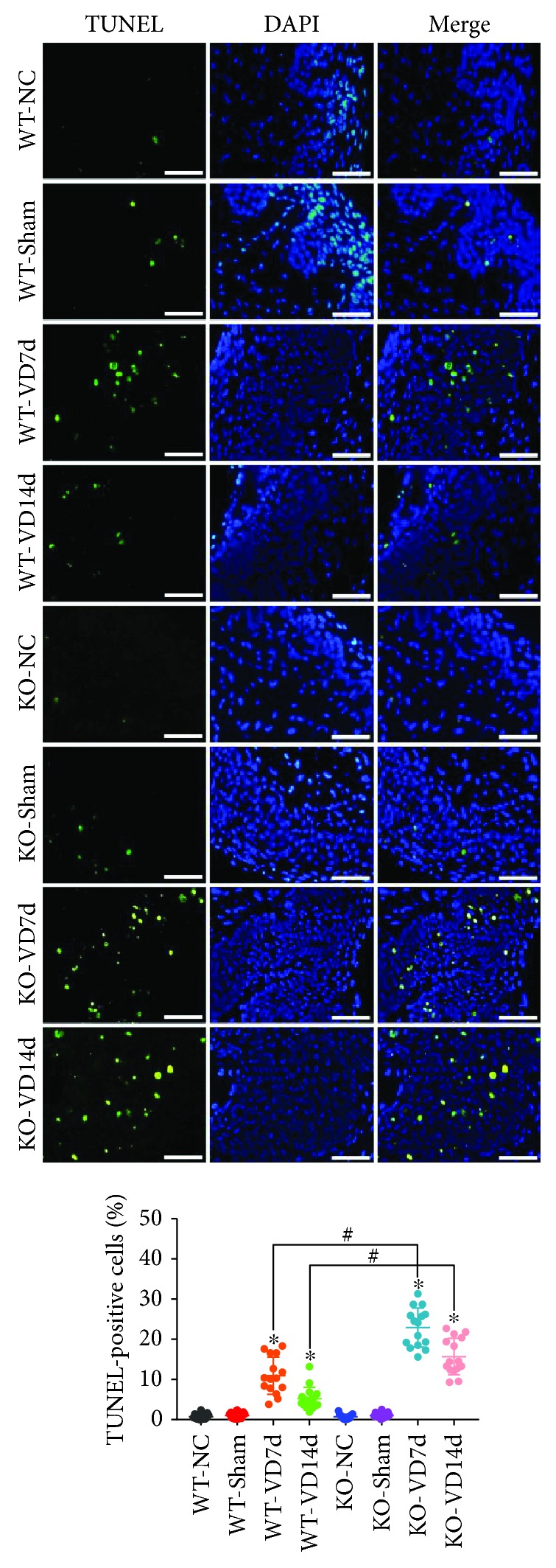
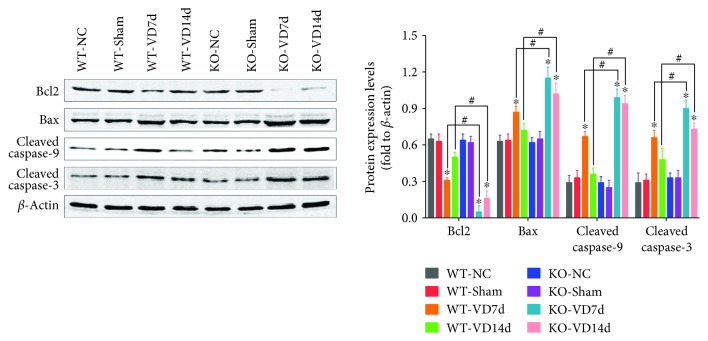
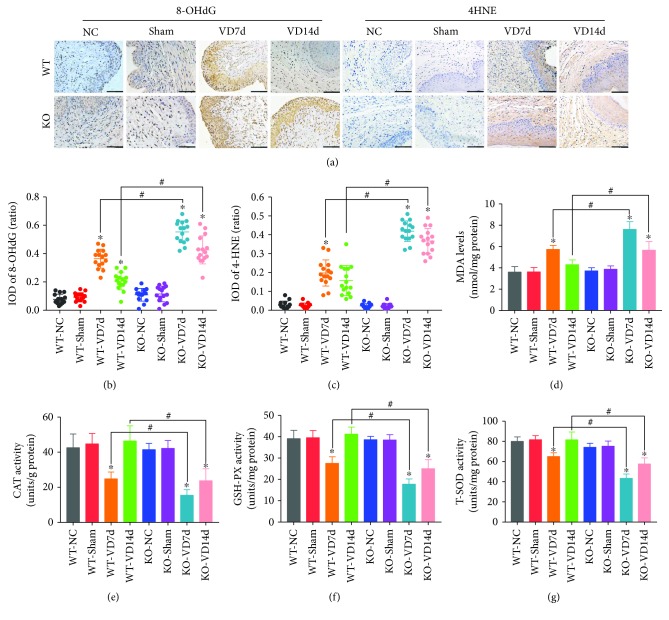
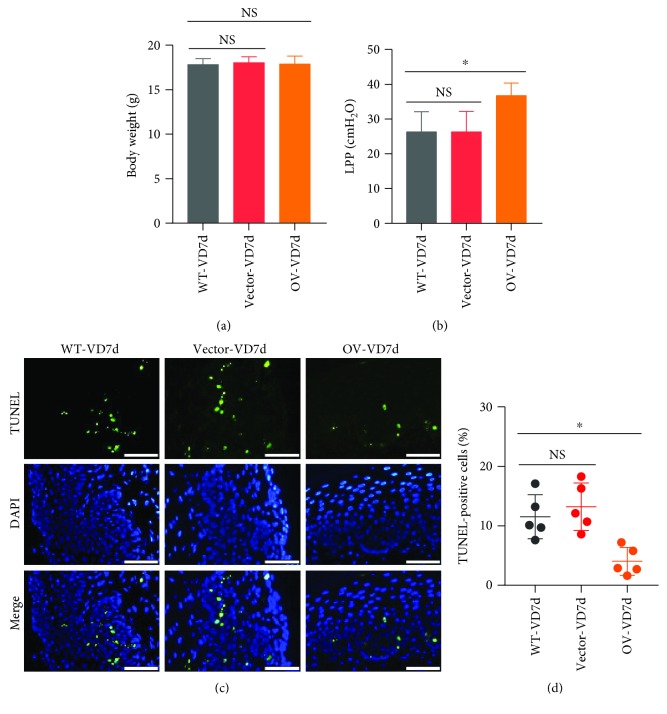
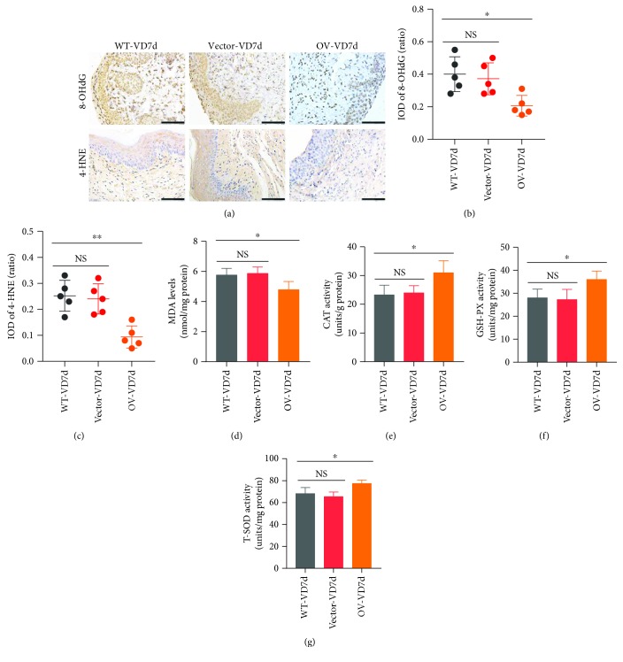
Apoptosis and oxidative damage are involved in the pathogenesis and progression of stress urinary incontinence (SUI). Our previous results indicate that cell apoptosis and oxidative damage increase in a mouse model of mechanical injury-induced SUI and in fibroblasts treated with excessive mechanical strain. Nuclear factor erythroid-2-related factor 2 (Nrf2) is a well-characterized global antioxidant gene inducer that can reduce oxidative damage and apoptosis. Therefore, we predicted that Nrf2 may have a protective role in mechanical trauma-induced SUI. To test this hypothesis, a mouse model of vaginal distension- (VD-) induced SUI was established. Leak point pressure (LPP); levels of apoptosis, apoptosis-related proteins, and peroxidation products; and the activities of antioxidative proteins in the anterior vaginal wall were measured in wild-type (Nfe2l2+/+) C57BL/6 mice and Nrf2-knockout mice (Nfe2l2-/-). The results showed that Nrf2 knockout aggravated VD-induced reduction in LPP, increase in cell apoptosis and peroxidation product levels, decrease in antioxidative protein activities, and alterations in apoptosis-related protein levels in the vaginal walls of mice. To further confirm the role of Nrf2 in mechanical trauma-induced apoptosis and SUI, VD was performed on mice overexpressing Nrf2 via in vivo transfection of LV-Nfe2l2. The results showed that Nrf2 overexpression significantly alleviated VD-induced abnormalities in the anterior vaginal wall. Taken together, our data suggested that Nrf2 is a potential protective factor in mechanical trauma-induced apoptosis in a mouse model of SUI. Antioxidative therapy may be a promising treatment for mechanical trauma-related SUI.


Figures

Figure 1
Figure 1
The body weights and LPP values of mice. (a) Body weights of mice in eight groups. (b) LPP values of mice in eight groups. P < 0.05 compared with the WT-VD group, # P < 0.05 between two groups. Every experiment was repeated 3 times. LPP: leak point pressure; VD: vaginal distension; WT-NC: nonoperated control group of wild-type mice; WT-Sham: sham-operated group of wild-type mice; WT-VD7d: VD group of wild-type mice and evaluated on day 7 after VD; WT-VD14d: VD group of wild-type mice and evaluated on day 14 after VD; KO-NC: nonoperated control group of Nrf2-knockout mice; KO-Sham: sham-operated group of Nrf2-knockout mice; KO-VD7d: VD group of Nrf2-knockout mice and evaluated on day 7 after VD; KO-VD14d: VD group of Nrf2-knockout mice and evaluated on day 14 after VD.
Figure 2
Figure 2
Apoptosis in the anterior vaginal wall of mice. TUNEL assay was used to detect apoptosis in the anterior wall of mice in eight groups. Green fluorescence is a positive signal for apoptosis. P < 0.05 compared with the WT-VD group, # P < 0.05 between two groups. Scale bars: 50 μm. Every experiment was repeated 3 times and representative figures are presented here. VD: vaginal distension; WT-NC: nonoperated control group of wild-type mice; WT-Sham: sham-operated group of wild-type mice; WT-VD7d: VD group of wild-type mice and evaluated on day 7 after VD; WT-VD14d: VD group of wild-type mice and evaluated on day 14 after VD; KO-NC: nonoperated control group of Nrf2-knockout mice; KO-Sham: sham-operated group of Nrf2-knockout mice; KO-VD7d: VD group of Nrf2-knockout mice and evaluated on day 7 after VD; KO-VD14d: VD group of Nrf2-knockout mice and evaluated on day 14 after VD.
Figure 3
Figure 3
Levels of apoptosis-related proteins in the anterior wall of mice. Western blot was used to determine the levels of apoptosis-related proteins in the anterior wall of mice in eight groups. P < 0.05 compared with the WT-VD group, # P < 0.05 between two groups. Every experiment was repeated 3 times, and representative figures were presented here. VD: vaginal distension; WT-NC: nonoperated control group of wild-type mice; WT-Sham: sham-operated group of wild-type mice; WT-VD7d: VD group of wild-type mice and evaluated on day 7 after VD; WT-VD14d: VD group of wild-type mice and evaluated on day 14 after VD; KO-NC: nonoperated control group of Nrf2-knockout mice; KO-Sham: sham-operated group of Nrf2-knockout mice; KO-VD7d: VD group of Nrf2-knockout mice and evaluated on day 7 after VD; KO-VD14d: VD group of Nrf2-knockout mice and evaluated on day 14 after VD.
Figure 4
Figure 4
Oxidative damage and antioxidative capacity in the anterior wall of mice. (a–c) Immunohistochemistry was used to detect the levels of 8-OHdG and 4-HNE in the anterior wall of mice in eight groups. Brown represents the positive signal. Scale bars: 100 μm. (d) The MDA measurement kit was used to detect the levels of MDA in the anterior wall of mice in eight groups. (e–g) CAT, GSH-PX, and T-SOD measurement kits were used to determine the activities of CAT, GSH-PX, and T-SOD, respectively, in the anterior wall of mice in eight groups. P < 0.05 compared with the WT-VD group, # P < 0.05 between two groups. Every experiment was repeated 3 times and representative figures are presented here. 8-OHdG: 8-oxo-2′-deoxyguanosine; 4-HNE: 4-hydroxynonenal; MDA: malondialdehyde; CAT: catalase; GSH-PX: glutathione peroxidase; T-SOD: total superoxide dismutase; VD: vaginal distension; WT-NC: nonoperated control group of wild-type mice; WT-Sham: sham-operated group of wild-type mice; WT-VD7d: VD group of wild-type mice and evaluated on day 7 after VD; WT-VD14d: VD group of wild-type mice and evaluated on day 14 after VD; KO-NC: nonoperated control group of Nrf2-knockout mice; KO-Sham: sham-operated group of Nrf2-knockout mice; KO-VD7d: VD group of Nrf2-knockout mice and evaluated on day 7 after VD; KO-VD14d: VD group of Nrf2-knockout mice and evaluated on day 14 after VD.
Figure 5
Figure 5
Effects of Nrf2 overexpression on mechanical trauma-induced LPP reduction and apoptosis. (a) Body weights of mice in three groups. (b) LPP values of mice in three groups. (c, d) TUNEL assay was used to detect the cell apoptosis in the anterior wall of mice in three groups. Green fluorescence is a positive signal for apoptosis. Scale bars: 50 μm. P < 0.05; NS: no significant difference. Every experiment was repeated 3 times and representative figures are presented here. LPP: leak point pressure; VD: vaginal distension; WT-VD7d: mice in the control group and evaluated on day 7 after VD; Vector-VD7d: mice transfected with LV vector and evaluated on day 7 after VD; Nrf2-OV: mice transfected with LV-Nfe2l2 for in vivo Nrf2 overexpression and evaluated on day 7 after VD.
Figure 6
Figure 6
Effects of Nrf2 overexpression on mechanical trauma-induced alterations of oxidative damage and antioxidative capacity. (a–c) Immunohistochemistry was used to detect the levels of 8-OHdG and 4-HNE in the anterior wall of mice in three groups. Brown represents the positive signal. Scale bars: 100 μm. (d) The MDA measurement kit was used to detect the levels of MDA in the anterior wall of mice in three groups. (e–g) CAT, GSH-PX, and T-SOD measurement kits were used to detect the activities of CAT, GSH-PX, and T-SOD, respectively, in the anterior wall of mice in three groups. P < 0.05; NS: no significant difference. Every experiment was repeated 3 times and representative figures are presented here. 8-OHdG: 8-oxo-2′-deoxyguanosine; 4-HNE: 4-hydroxynonenal; MDA: malondialdehyde; CAT: catalase; GSH-PX: glutathione peroxidase; T-SOD: total superoxide dismutase; VD: vaginal distension; WT-VD7d: mice in control group and evaluated on day 7 after VD; Vector-VD7d: mice transfected with LV vector and evaluated on day 7 after VD; Nrf2-OV: mice transfected with LV-Nfe2l2 for in vivo Nrf2 overexpression and evaluated on day 7 after VD.

References

    1. Lavelle E. S., Zyczynski H. M. Stress urinary incontinence: comparative efficacy trials. Obstetrics and Gynecology Clinics of North America. 2016;43(1):45–57. doi: 10.1016/j.ogc.2015.10.009. - DOI - PubMed
    1. Syan R., Brucker B. M. Guideline of guidelines: urinary incontinence. BJU International. 2016;117(1):20–33. doi: 10.1111/bju.13187. - DOI - PubMed
    1. Barber M. D., Maher C. Epidemiology and outcome assessment of pelvic organ prolapse. International Urogynecology Journal. 2013;24(11):1783–1790. doi: 10.1007/s00192-013-2169-9. - DOI - PubMed
    1. Zhu L., Lang J., Liu C., Han S., Huang J., Li X. The epidemiological study of women with urinary incontinence and risk factors for stress urinary incontinence in China. Menopause. 2009;16(4):831–836. doi: 10.1097/gme.0b013e3181967b5d. - DOI - PubMed
    1. DeLancey J. O. L. Fascial and muscular abnormalities in women with urethral hypermobility and anterior vaginal wall prolapse. American Journal of Obstetrics and Gynecology. 2002;187(1):93–98. doi: 10.1067/mob.2002.125733. - DOI - PubMed

Substances