

Perturbed Biochemical Pathways and Associated Oxidative Stress Lead to Vascular Dysfunctions in Diabetic Retinopathy
- PMID: 30962865
- PMCID: PMC6431380
- DOI: 10.1155/2019/8458472
Perturbed Biochemical Pathways and Associated Oxidative Stress Lead to Vascular Dysfunctions in Diabetic Retinopathy
Abstract
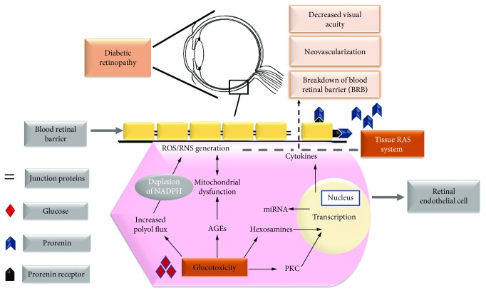
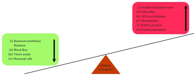
Diabetic retinopathy (DR) is a vascular insult that accompanies the hyperglycemic state. Retinal vasculature holds a pivotal role in maintaining the integrity of the retina, and any alteration to retinal vasculature affects retinal functions. The blood retinal barrier, a prerequisite to vision acuity, is most susceptible to damage during the progression of DR. This is a consequence of impaired biochemical pathways such as the polyol, advanced end glycation products (AGE), hexosamine, protein kinase C (PKC), and tissue renin-angiotensin system (RAS) pathways. Moreover, the role of histone modification and altered miRNA expression is also emerging as a major contributor. Epigenetic changes create a link between altered protein function and redox status of retinal cells, creating a state of metabolic memory. Although various biochemical pathways underlie the etiology of DR, the major insult to the retina is due to oxidative stress, a unifying factor of altered biochemical pathways. This review primarily focuses on the critical biochemical pathways altered in DR leading to vascular dysfunctions and discusses antioxidants as plausible treatment strategies.
Figures
References
Publication types
MeSH terms
LinkOut - more resources
Full Text Sources
Medical
