Hypoglossal Nerve Palsy as a Cause of Severe Dysphagia along with the Oropharyngeal Stenosis due to Occipitocervical Kyphosis
- PMID: 30963014
- PMCID: PMC6431363
- DOI: 10.1155/2019/7982847
Hypoglossal Nerve Palsy as a Cause of Severe Dysphagia along with the Oropharyngeal Stenosis due to Occipitocervical Kyphosis
Abstract
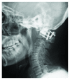
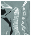
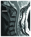
Hypoglossal nerve palsy (HNP) is a potential cause of dysphagia. A 66-year-old man presented to our hospital with dysphagia and neck pain. One year prior to his first visit, he had been diagnosed with upper cervical tuberculosis and had undergone posterior C1-2 fixation. The physical examination led to the diagnosis of dysphagia with HNP, and he had severe weight loss. Radiographic examination revealed that the O-C kyphosis had been exacerbated and that the deformity was likely the primary cause of HNP. To restore the swallowing function, O-C fusion surgery was performed. Postoperatively, the patient showed immediate improvement of dysphagia with gradual recovery of hypoglossal nerve function. In the last follow-up evaluation, swallowing function was confirmed with no signs of HNP. Our results indicate that HNP could be more prevalent in cases with severe cervical kyphosis, being underdiagnosed due to the more apparent signs of the oropharyngeal narrowing.
Figures


Similar articles
-
Microvascular decompression for hypoglossal nerve palsy secondary to vertebral artery compression: A case report and review of the literature.Surg Neurol Int. 2017 May 10;8:74. doi: 10.4103/sni.sni_42_17. eCollection 2017. Surg Neurol Int. 2017. PMID: 28584677 Free PMC article.
-
Ipsilateral hypoglossal nerve palsy following left hemithyroidectomy: Case report and review of literature.Int J Surg Case Rep. 2018;51:5-7. doi: 10.1016/j.ijscr.2018.07.044. Epub 2018 Aug 11. Int J Surg Case Rep. 2018. PMID: 30121396 Free PMC article.
-
Hypoglossal Nerve Palsy Following Cervical Spine Surgery-Two Case Reports and a Systematic Review of the Literature.Brain Sci. 2025 Feb 27;15(3):256. doi: 10.3390/brainsci15030256. Brain Sci. 2025. PMID: 40149777 Free PMC article. Review.
-
Hypoglossal nerve palsy after posterior screw placement on the C-1 lateral mass. Case report.J Neurosurg Spine. 2006 Jul;5(1):83-5. doi: 10.3171/spi.2006.5.1.83. J Neurosurg Spine. 2006. PMID: 16850964
-
Idiopathic Isolated Unilateral Hypoglossal Nerve Palsy: A Report of 2 Cases and Review of the Literature.J Oral Maxillofac Surg. 2018 Jul;76(7):1454-1459. doi: 10.1016/j.joms.2018.01.019. Epub 2018 Feb 2. J Oral Maxillofac Surg. 2018. PMID: 29452069 Review.
Cited by
-
Analysis of risk factors for postoperative dysphagia after C1-2 fusion.Front Surg. 2022 Oct 13;9:977500. doi: 10.3389/fsurg.2022.977500. eCollection 2022. Front Surg. 2022. PMID: 36311942 Free PMC article.
-
Tongue atrophy as a neurological finding in hereditary polyneuropathy in Alaskan malamutes.J Vet Intern Med. 2022 Mar;36(2):672-678. doi: 10.1111/jvim.16351. Epub 2022 Jan 12. J Vet Intern Med. 2022. PMID: 35019187 Free PMC article.
References
Publication types
LinkOut - more resources
Full Text Sources

