. 2019 Nov 25;40(11):1415-1426.
doi: 10.1093/carcin/bgz070.

β-hCG-induced mutant BRCA1 ignites drug resistance in susceptible breast tissue

Affiliations

β-hCG-induced mutant BRCA1 ignites drug resistance in susceptible breast tissue

Satheesh Kumar Sengodan et al. Carcinogenesis. .

Abstract

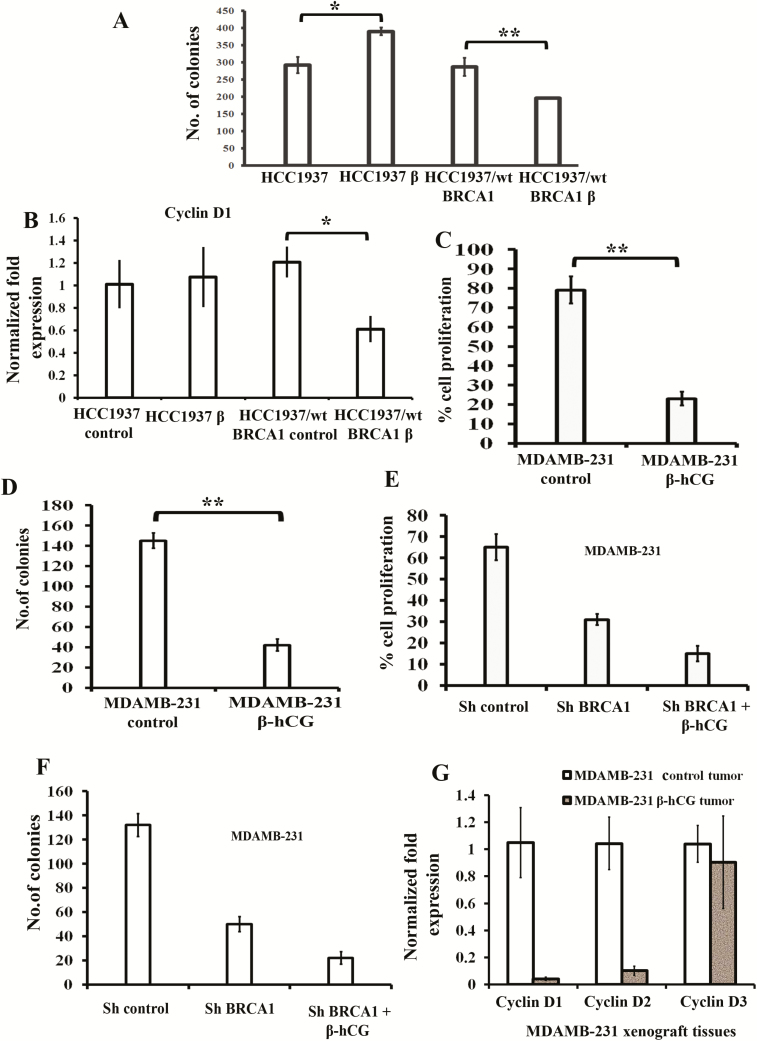
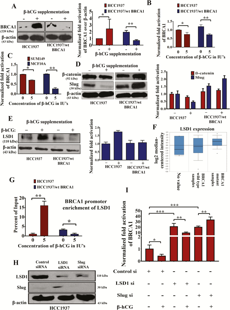
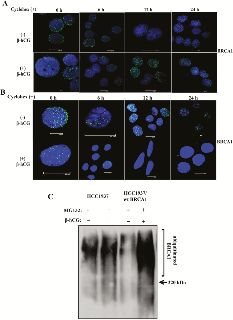
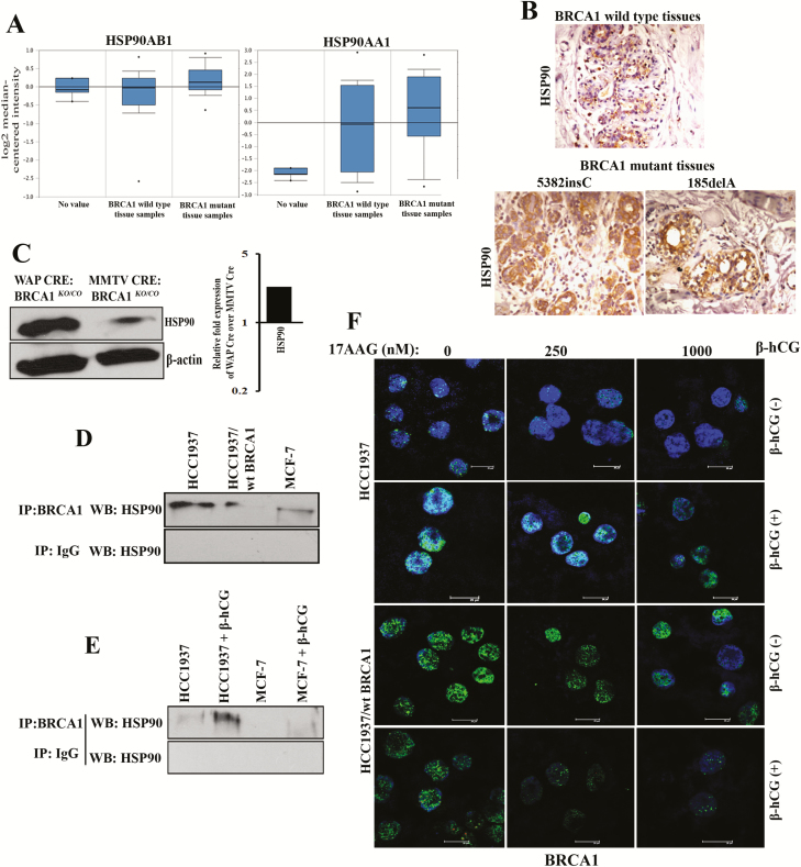
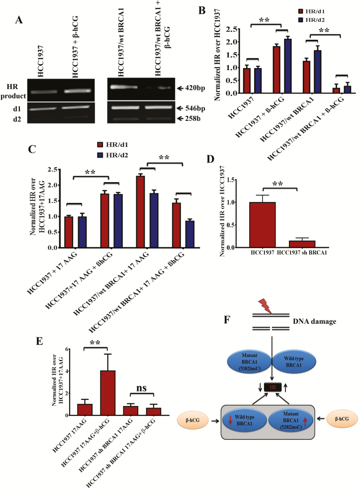
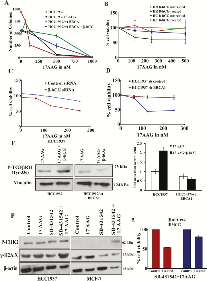
β-hCG expression in breast cancer is highly controversial with reports supporting both protective and tumorigenic effects. It has also been reported that risk of breast cancer at an early age is increased with full-term pregnancies if a woman is a BRCA1 mutation carrier. We have already demonstrated that BRCA1-defective cells express high levels of β-hCG and that when BRCA1 is restored, β-hCG level is reduced. Also, BRCA1 can bind to the promoter and reduce the levels of β-hCG. β-hCG induces tumorigenicity in BRCA1-defective cells by directly binding to TGFBRII and induces TGFBRII-mediated cell proliferation. In this study, we analyzed the mechanism of action of β-hCG on BRCA1 expression and its influence on drug sensitivity in breast cancer cells. We demonstrate that β-hCG induces mutant BRCA1 protein expression in BRCA1 mutant cells; however, in BRCA1 wild-type cells, β-hCG reduced wild-type BRCA1 protein expression. Transcriptionally, β-hCG could induce Slug/LSD1-mediated repression of wild-type and mutant BRCA1 messenger RNA levels. However, β-hCG induces HSP90-mediated stabilization of mutant BRCA1 and hence the overexpression of mutant BRCA1 protein, resulting in partial restoration of homologous recombination repair of damaged DNA. This contributes to drug resistance to HSP90 inhibitor 17AAG in BRCA1-defective cancer cells. A combination of HSP90 inhibitor and TGFBRII inhibitor has shown to sensitize β-hCG expressing BRCA1-defective breast cancers to cell death. Targeting the β-hCG-HSP90-TGFBRII axis could prove an effective treatment strategy for BRCA1-mutated breast tumors.


Figures

Figure 1.
Figure 1.
β-hCG induces proliferation in BRCA1-mutated but reduces proliferation in BRCA1 wild-type breast cancer cells. (A) Analysis of cell proliferation by colony formation assay in β-hCG overexpressing HCC1937 (HCC1937 β) and HCC1937/wt BRCA1 (HCC1937/wt BRCA1 β). (B) RT-qPCR analysis of Cyclin D1 from β-hCG overexpressing HCC1937 (HCC1937 β) and HCC1937/wt BRCA1 (HCC1937/wt BRCA1 β). Cell proliferation assay of stable β-hCG overexpressing MDAMB-231 cells using (C) MTS and (D) colony formation assay. Cell proliferation assay of stable BRCA1 silenced and/or BRCA1 silenced with β-hCG overexpressing MDAMB-231 using (E) MTS assay and (F) colony formation assay. (G) Quantitative Real Time PCR (qRT-PCR) analysis of Cyclin D1, Cyclin D2 and Cyclin D3 from xenograft tumor induced by Control MDAMB-231 and/or β-hCG overexpressing MDAMB-231 cells. All the experiments are conducted in triplicates. All error bars in the graphs represent SD. *P < 0.05, **P < 0.01, ***P < 0.001.
Figure 2.
Figure 2.
β-hCG induces mutant but not wild-type BRCA1. (A) Western blot analysis of BRCA1 upon supplementation of 5 IU of β-hCG for 24 h in HCC1937 and HCC1937/wt BRCA1 cells. Quantification of the western blot on the right panel. β-actin was used as an endogenous control. (B) Quantitative Real Time PCR (qRT-PCR) analysis of BRCA1 expression upon supplementation of 5 IU of β-hCG for 24 h in HCC1937 and HCC1937/wt BRCA1. (C) qRT-PCR analysis of BRCA1 expression upon supplementation of 5 IU of β-hCG for 24 h in SUM149 and MCF10A. (D) Western blot analysis of β-catenin and Slug upon supplementation of 5 IU of β-hCG for 24 h in HCC1937 and HCC1937/wt BRCA1 cells. Quantitation was given on right panel. (E) Western blot analysis of LSD1 upon supplementation of 5 IU of β-hCG for 24 h in HCC1937 and HCC1937/wt BRCA1. Quantitation was given on right panel. (F) TCGA dataset analysis of LSD1 in BRCA1 wild-type and BRCA1 mutant human breast cancer tissue samples (Waddell’s dataset) using Oncomine. (G) qRT-PCR amplification of BRCA1 promoter immunoprecipitated with anti-LSD1 antibody from HCC1937 and HCC1937/wt BRCA1 supplemented with and/or without β-hCG. Normal chromatin fragments were used as input. (H) Western blot confirmation of silencing of LSD1 and Slug in HCC1937. (I) RT-qPCR analysis of BRCA1 in control, LSD1 and Slug-silenced HCC1937 in the presence and/or absence of β-hCG. Full blots of cropped images are included in supplementary information. *P < 0.05, **P < 0.01, ***P < 0.001.
Figure 3.
Figure 3.
β-hCG stabilizes mutant but not wild-type BRCA1. (A) Expression BRCA1 upon treating with 100 μM of cycloheximide (Cyclohex) over a time-period of up to 24 h in the presence (+) or in the absence (−) of β-hCG in HCC1937. (B) Similar experiment was performed to analyze expression of BRCA1 in HCC1937/wt BRCA1. Scale bars, 20 μm. Blue color in the immunofluorescence images represents 4′,6-diamidino-2-phenylindole, which stains the nucleus, whereas the green color indicates the staining for BRCA1 protein. (C) Western blot analysis of ubiquitin from β-hCG-supplemented HCC1937 and HCC1937/wt BRCA1 cells immunoprecipitated with anti-BRCA1 antibody. Proteasome-mediated degradation of protein from HCC1937 and HCC1937/wt BRCA1 was blocked using MG132. Full blots of cropped images are included in supplementary information.
Figure 4.
Figure 4.
Presence of β-hCG results in the stabilization of BRCA1 even in the presence of HSP90 inhibitor. (A) TCGA dataset analysis of HSP90AB1 and HSP90AA1 in BRCA1 wild-type and BRCA1 mutant human breast cancer tissue samples (Waddell’s dataset) using Oncomine. (B) Immunohistochemical analysis of HSP90 in BRCA1 wild-type and BRCA1 mutant (5382insC and 185delA) human breast cancer tissues. (C) Western blot analysis of HSP90 from WAP-Cre; BRCA1KO/CO and MMTV-Cre; BRCA1KO/CO mouse tumor samples. Quantitation of the western blot is given on the right panel. (D) Western blot analysis of HSP90 immunoprecipitated with BRCA1 or IgG antibody in HCC1937, HCC1937/wt BRCA1 and MCF-7. (E) Western blot analysis of HSP90 immunoprecipitated with BRCA1 or IgG antibody in HCC1937 and MCF-7 supplemented with β-hCG. (F) Immunofluorescence for BRCA1 upon treating with 17AAG for a period of 24 h in the presence (+) or in the absence (−) of β-hCG in HCC1937 and HCC1937/wt BRCA1. Scale bars, 20 μm. Blue color in the immunofluorescence images represents 4′,6-diamidino-2-phenylindole, which stains the nucleus, whereas the green color indicates the staining for BRCA1 protein. Full blots of cropped images are included in supplementary information.
Figure 5.
Figure 5.
β-hCG induces HR in BRCA1 mutant but not in wild-type cells. (A) HR assay showing the level of recombination in HCC1937 and HCC1937/wt BRCA1 upon supplementation of β-hCG. d1 and d2 plasmid were taken as internal control. The d1 and d2 were obtained as 546bp and 258bp products whereas recombination product (HR) was obtained at 420bp. (B) HR levels were normalized to HCC1937 and quantitation of HR was given on right panel. (C) Quantitative Real Time PCR (qRT-PCR) analysis showing the level of HR in HCC1937 and HCC1937/wt BRCA1 cells treated with 17AAG in the presence or absence of β-hCG. HR levels were normalized to HCC1937 + 17AAG. (D) qRT-PCR analysis showing the level of HR in HCC1937 and mutant BRCA1 stably silenced HCC1937 cells (HCC1937 sh BRCA1). (E) qRT-PCR analysis showing the level of HR in HCC1937 and mutant BRCA1 stably silenced HCC1937 cells (HCC1937 sh BRCA1) treated with 17AAG in the presence or absence of β-hCG. (F) Pictorial depiction of HR in BRCA1 mutant and wild-type cells with respect to β-hCG. Full blots of cropped images are included in supplementary information. **P < 0.01.
Figure 6.
Figure 6.
β-hCG induces drug resistance in BRCA1 mutant cells. (A) Colony-forming ability (adherent colonies) of HCC1937 and HCC1937/wt BRCA1 in the presence or in the absence of 5 IU of β-hCG after treating with indicated doses of 17AAG for 72 h. (B) Cell proliferation of HCC1937 and HCC1937/wt BRCA1 cells treated with increasing concentrations of 17AAG for 96 h in the presence or in the absence of β-hCG as assessed by MTS assay. (C) Cell proliferation of β-hCG-silenced HCC1937 cells treated with increasing concentrations of 17AAG for 72 h as assessed by MTS assay. (D) Cell proliferation of BRCA1 stably silenced HCC1937 cells treated with increasing concentrations of 17AAG for 72 h as assessed by MTS assay. (E) The level of phosphorylation of TGFBRII in HCC1937 and HCC1937/wt BRCA1cells treated with and/or without 17AAG in the presence or absence of β-hCG. (F) Western blot analysis of P-CHK2 and γ-H2A.X in HCC1937 and MCF7 treated with 17AAG or SB-431542 or combination of both. (G) Cell proliferation of HCC1937 and MCF7 treated with combination of 17AAG and SB-431542 as assessed by MTS assay. Full blots of cropped images are included in supplementary information.

References

    1. Russo J., et al. (2005) The protective role of pregnancy in breast cancer. Breast Cancer Res., 7, 131–142. - PMC - PubMed
    1. Meier-Abt F., et al. (2014) How pregnancy at early age protects against breast cancer. Trends Mol. Med., 20, 143–153. - PubMed
    1. Meier-Abt F., et al. (2013) Parity induces differentiation and reduces Wnt/Notch signaling ratio and proliferation potential of basal stem/progenitor cells isolated from mouse mammary epithelium. Breast Cancer Res., 15, R36. - PMC - PubMed
    1. Jernström H., et al. (1999) Pregnancy and risk of early breast cancer in carriers of BRCA1 and BRCA2. Lancet, 354, 1846–1850. - PubMed
    1. Andrieu N., et al. ; EMBRACE; GENEPSO; GEO-HEBON; IBCCS Collaborators Group (2006) Pregnancies, breast-feeding, and breast cancer risk in the International BRCA1/2 Carrier Cohort Study (IBCCS). J. Natl. Cancer Inst., 98, 535–544. - PMC - PubMed

MeSH terms

Substances

LinkOut - more resources