Review
. 2020 Apr;139(4):625-641.
doi: 10.1007/s00401-019-02002-2. Epub 2019 Apr 8.

An update on the central nervous system manifestations of neurofibromatosis type 1

Affiliations
Review

An update on the central nervous system manifestations of neurofibromatosis type 1

J Stephen Nix et al. Acta Neuropathol. 2020 Apr.

Abstract

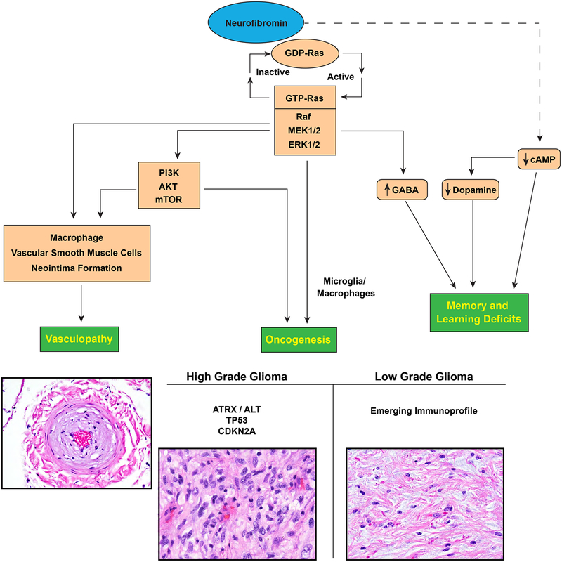
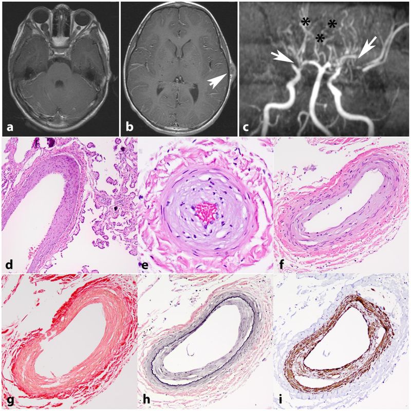
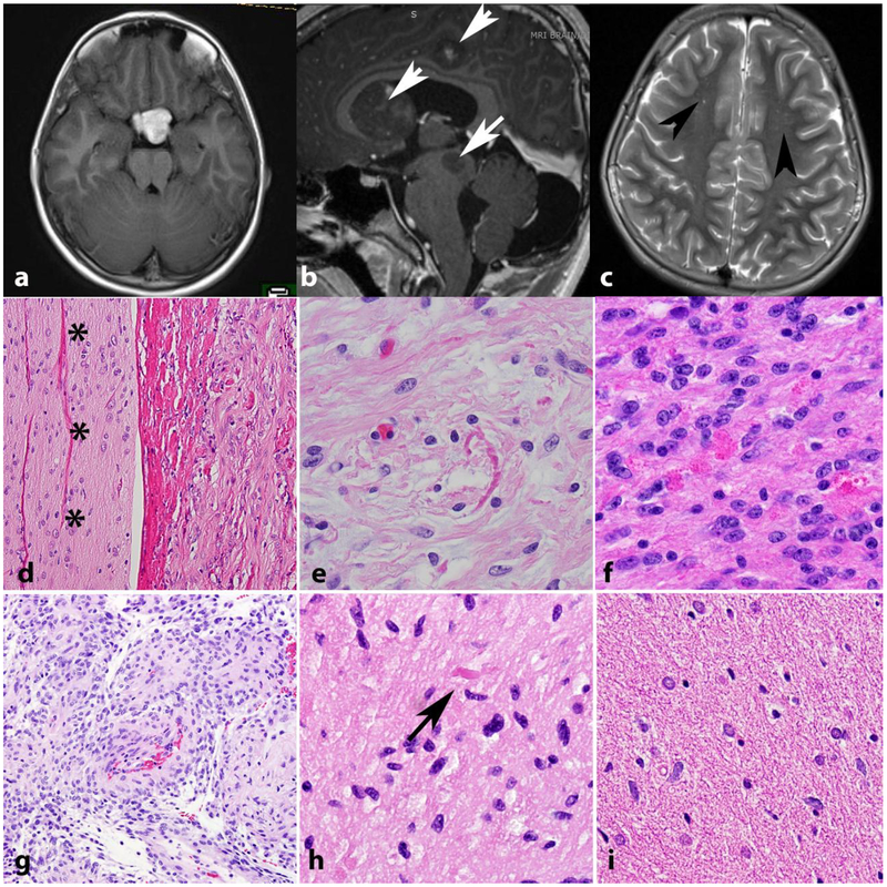
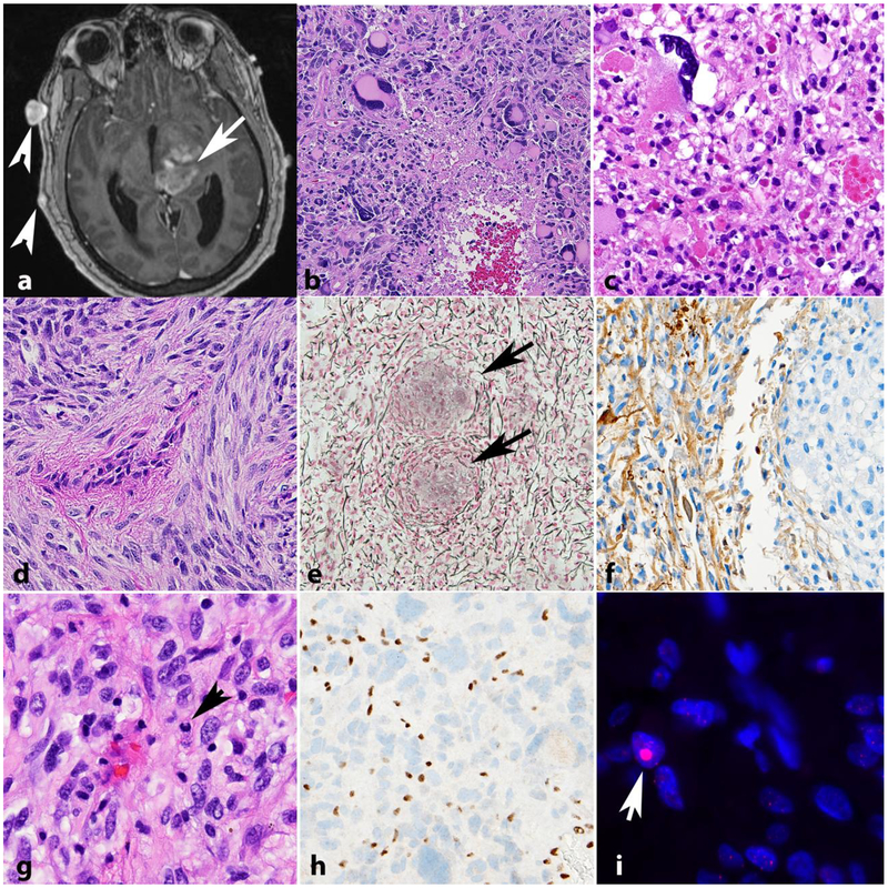
Neurofibromatosis 1 (NF1) is an autosomal dominant genetic disorder that presents with variable phenotypes as a result of mutations in the neurofibromatosis type 1 (NF1) gene and subsequently, abnormal function of the protein product, neurofibromin. Patients with NF1 are at increased risk for central nervous system (CNS) manifestations including structural, functional, and neoplastic disease. The mechanisms underlying the varied manifestations of NF1 are incompletely understood, but the loss of functional neurofibromin, resulting in sustained activation of the oncoprotein RAS, is responsible for tumorigenesis throughout the body, including the CNS. Much of our understanding of NF1-related CNS manifestations is from a combination of data from animal models and natural history studies of people with NF1 and CNS disease. Data from animal models suggest the importance of both Nf1 mutations and somatic genetic alterations, such as Tp53 loss, for development of neoplasms, as well as the role of the timing of the acquisition of such alterations on the variability of CNS manifestations. A variety of non-neoplastic structural (macrocephaly, hydrocephalus, aqueductal stenosis, and vasculopathy) and functional (epilepsy, impaired cognition, attention deficits, and autism spectrum disorder) abnormalities occur with variable frequency in individuals with NF1. In addition, there is increasing evidence that similar appearing CNS neoplasms in people with and without the NF1 syndrome are due to distinct oncogenic pathways. Gliomas in people with NF1 show alterations in the RAS/MAPK pathway, generally in the absence of BRAF alterations (common to sporadic pilocytic astrocytomas) or IDH or histone H3 mutations (common to diffuse gliomas subsets). A subset of low-grade astrocytomas in these patients remain difficult to classify using standard criteria, and occasionally demonstrate morphologic features resembling subependymal giant cell astrocytomas that afflict patients with tuberous sclerosis complex ("SEGA-like astrocytomas"). There is also emerging evidence that NF1-associated high-grade astrocytomas have frequent co-existing alterations such as ATRX mutations and an alternative lengthening of telomeres (ALT) phenotype responsible for unique biologic properties. Ongoing efforts are seeking to improve diagnostic accuracy for CNS neoplasms in the setting of NF1 versus sporadic tumors. In addition, MEK inhibitors, which act on the RAS/MAPK pathway, continue to be studied as rational targets for the treatment of NF1-associated tumors, including CNS tumors.

Keywords: Brain tumor; Glioma; Hydrocephalus; Neurofibromatosis; Neurofibromin; Seizure; Vasculopathy.


Figures

Figure 1.
Figure 1.
CNS manifestations resulting from neurofibromin loss and aberrant signaling pathways.
Figure 2.
Figure 2.. Intracranial vasculopathy in NF1.
Axial T1 weighted image with contrast in a 15-year-old boy with NF1 shows bilateral optic nerve gliomas, larger on the right (a). Axial T1 weighted image with contrast also shows a left frontoparietal scalp soft tissue lesion representing a cutaneous neurofibroma (arrow) (b). MR angiography of the circle of Willis shows stenosis of the distal internal carotid arteries, with no flow in the right middle cerebral artery and severe narrowing of the left middle cerebral artery (arrows). Many collateralized vessels in a moya-moya pattern are seen (asterisks) (c). Histologic sections from a 21 year-old patient with NF1 demonstrating intimal thickening involving vessels in the choroid plexus (d) and leptomeninges (e,f). Picrosirius red stain demonstrates normal mural collagen fiber architecture (g). VVG highlights intimal hyperplasia central to internal elastic lamina (black)(h) and SMA immunostain shows positivity in many of these cells (i).
Figure 3.
Figure 3.. Low grade astrocytomas in NF1.
The optic pathways, including the optic chiasm are favored sites for low grade astrocytomas in NF1. Axial T1 weighted MR image in a 41-year-old woman with NF1 demonstrates a bulky mass involving the optic chiasm (a) that histologically proved to be a pilocytic astrocytoma. Multicentricity (arrows) of pilocytic astrocytomas is a feature of NF1 (arrows) (b). “Unidentified bright objects” represent hyperintensities on MRI that do not grow and frequently regress over time (arrowheads). They are frequent in individuals with NF1 and should not be misinterpreted as tumors (Axial T2-weighted image) (c). Pilocytic astrocytomas involving the optic nerve (asterisks) (optic nerve glioma) frequently extend into the subarachnoid space (d). Most gliomas in individuals with NF1 are pilocytic astrocytomas, characterized by frequent Rosenthal fibers (e) and eosinophilic granular bodies (f). The pilomyxoid variant of pilocytic astrocytoma also may develop in individuals with NF1 (g). Low grade astrocytomas that are difficult to classify, usually having infiltrative features but with occasional piloid features such as rare Rosenthal fibers (arrow) are also relatively frequent (h). Diffuse astrocytomas also occur in individuals with NF1 and resemble sporadic diffuse astrocytomas, particularly single cell infiltration and lack of Rosenthal fibers(i).
Figure 4.
Figure 4.. High grade astrocytomas in NF1.
High grade astrocytoma forming a deep contrast enhancing mass (arrow). Other manifestations of NF1 in this patient included neurofibromas of the scalp (arrowheads) (axial T1-weighted post-contrast image)(a). The histology of high grade astrocytomas in individuals with NF1 is also variable, and includes giant cell glioblastoma (b) and anaplastic pleomorphic xanthoastrocytoma (c). NF1 associated gliosarcoma (d) with biphasic components highlighted by reticulin special stain, including islands of reticulin poor glioma cells (arrows) surrounded by reticulin rich sarcomatous areas (e). GFAP immunoreactivity in this tumor is limited to glial areas (left) and is negative in the pleomorphic sarcomatous component (right)(f). Anaplastic astrocytoma in NF1 patient with mitotic activity (arrow)(g), ATRX expression loss by immunohistochemistry (h) and large foci in telomeric FISH (arrow) consistent with alternative lengthening of telomeres (i).
Figure 5.
Figure 5.. Glioneuronal tumors in NF1.
Gangliogliomas centered in the conus (a,b,d,e) and occipital cortex (c,f) in individuals with NF1. CD34 expression may be seen (e) as in sporadic gangliogliomas. Dysembryoplastic neuroepithelial tumor forming mucoid cortical nodules (arrows)(g) and containing floating neurons on high power (h). Rosette forming glioneuronal tumors also may occur in individuals with NF1, often outside the posterior fossa (i).
Figure 6.
Figure 6.. SEGA-like astrocytoma.
Neoplasms resembling subependymal giant cell astrocytomas (SEGA) typical of tuberous sclerosis occur in a subset of individuals with NF1, although they tend to be hemispheric (a, b,c). As SEGA, they express glial markers (GFAP, d; OLIG2, e) and neuronal markers (synaptophysin, f). SEGA-like astrocytoma in NF1 patient (g) with intact ATRX expression (h) but large telomere foci (red) colocalizing with Promyelocytic bodies (green)(arrow) consistent with alternative lengthening of telomeres (i).
Figure 7.
Figure 7.. Sporadic pilocytic astrocytoma with anaplasia and NF1 pathogenic gene variant.
Axial T1-weighted MRI demonstrating a thalamic mass with heterogeneous enhancement (arrow), as well as intraventricular nodules consistent with subependymal spread (arrowheads) (a). First biopsy demonstrated a pilocytic astrocytoma (b). Recurrence in the absence of treatment showed a cellular glial neoplasm with brisk mitotic activity consistent with anaplastic progression (c). Next generation sequencing demonstrated a somatic truncating NF1 p.R816* variant with a variant allele frequency (VAF) of 38.5 (c). No other significant gene sequencing variants were identified.

References

    1. Anastasaki C, Gutmann DH (2014) Neuronal NF1/RAS regulation of cyclic AMP requires atypical PKC activation. Hum Mol Genet 23:6712–6721. doi:10.1093/hmg/ddu389 - DOI - PMC - PubMed
    1. Appin CL, Brat DJ (2014) Molecular Genetics of Gliomas. Cancer J 20:66–72. doi:10.1097/ppo.0000000000000020 - DOI - PubMed
    1. Avery RA, Hwang EI, Ishikawa H, Acosta MT, Hutcheson KA, Santos D, Zand DJ, Kilburn LB, Rosenbaum KN, Rood BR, Schuman JS, Packer RJ (2014) Handheld optical coherence tomography during sedation in young children with optic pathway gliomas. JAMA Ophthalmol 132:265–271. doi:10.1001/jamaophthalmol.2013.7649 - DOI - PMC - PubMed
    1. Bajaj A, Li QF, Zheng Q, Pumiglia K (2012) Loss of NF1 expression in human endothelial cells promotes autonomous proliferation and altered vascular morphogenesis. PLoS One 7:e49222. doi:10.1371/journal.pone.0049222 - DOI - PMC - PubMed
    1. Bajenaru ML, Donahoe J, Corral T, Reilly KM, Brophy S, Pellicer A, Gutmann DH (2001) Neurofibromatosis 1 (NF1) heterozygosity results in a cell-autonomous growth advantage for astrocytes. Glia 33:314–323. doi:10.1002/1098-1136(20010315)33:4<314::AIDGLIA1030>3.0.CO;2-Q - DOI - PubMed

Publication types

MeSH terms