

Acute appendicitis associated with the presence of schistosome eggs in a sailor: a case report
- PMID: 30963331
- PMCID: PMC6453986
- DOI: 10.1186/s40792-019-0615-8
Acute appendicitis associated with the presence of schistosome eggs in a sailor: a case report
Abstract
Background: Schistosomiasis is prevalent in tropical and subtropical areas and rarely reported in developed countries. Schistosomiasis often occurs as a chronic illness, which can cause liver and intestinal damage. Appendicitis is an unusual complication of schistosomiasis. We herein present a case of acute appendicitis associated with the presence of schistosome eggs in a sailor from the Philippines.
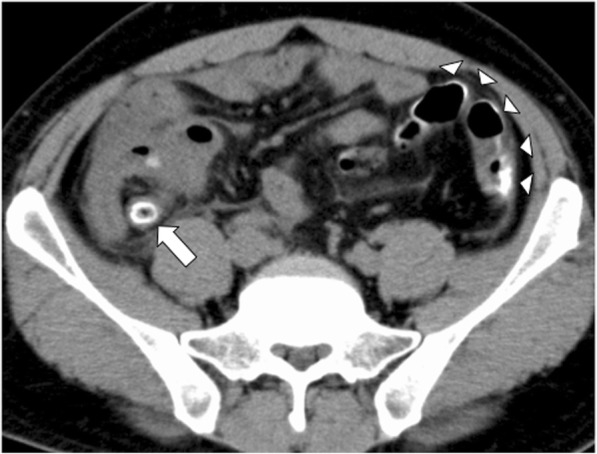
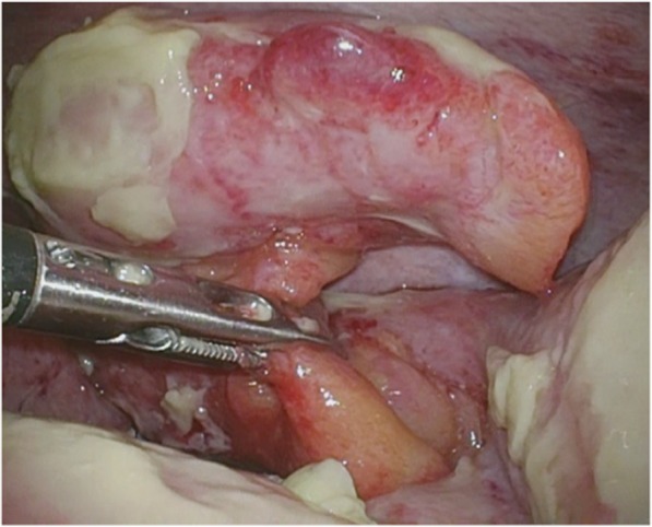
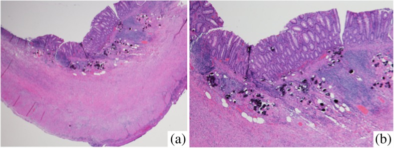
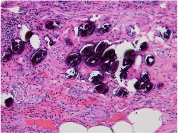
Case presentation: A 34-year-old Filipino man who worked as a sailor presented to our hospital with a 2-day history of acute right lower quadrant abdominal pain and fever. A physical examination revealed right lower quadrant abdominal pain with signs of peritoneal irritation, including rebound tenderness. Computed tomography revealed thickening of the appendix associated with mural calcification and fluid collection around the cecum. Based on these findings, the preoperative diagnosis was acute appendicitis. Laparoscopic appendectomy was performed. Swelling of the appendix and contaminated ascites were observed intraoperatively, but there was no evidence of appendiceal perforation. A histopathological examination showed inflammation of the appendix wall and numerous ovoid bodies present within the submucosa, many of which were calcified. Severe infiltration of lymphocytes and fibrosis were recognized around the oval bodies. The numerous oval bodies were morphologically consistent with schistosomiasis. The final diagnosis was acute phlegmonous appendicitis associated with the presence of schistosome eggs. We examined the patient for signs of adult worm activity, but the results of stool ova and parasite examinations performed twice were negative. He was discharged and returned to his country on postoperative day 9.
Conclusions: The incidence of schistosomal appendicitis, which is seldom reported in developed countries, is expected to increase in Japan in the near future. Clinicians should suspect schistosome eggs as a cause of acute appendicitis in patients who have emigrated from or are traveling from endemic areas, and when mural calcification of the appendix is observed on imaging.
Keywords: Appendicitis; Appendix; Mural calcification; Schistosomal appendicitis; Schistosomiasis.
Conflict of interest statement
Ethics approval and consent to participate
Not applicable.
Consent for publication
Written informed consent was obtained from the patient for the publication of this case report and any accompanying images.
Competing interests
The authors declare that they have no competing interests.
Publisher’s Note
Springer Nature remains neutral with regard to jurisdictional claims in published maps and institutional affiliations.
Figures
References
-
- WHO. Schistosomiasis fact sheet of 20 February 2018. Available online: http://www.who.int/news-room/fact-sheets/detail/schistosomiasis (Accessed 20 Aug 2018).
LinkOut - more resources
Full Text Sources
