. 2019 Oct;70(4):1231-1245.
doi: 10.1002/hep.30646. Epub 2019 May 24.

A Frizzled-Like Cysteine-Rich Domain in Glypican-3 Mediates Wnt Binding and Regulates Hepatocellular Carcinoma Tumor Growth in Mice

Affiliations

A Frizzled-Like Cysteine-Rich Domain in Glypican-3 Mediates Wnt Binding and Regulates Hepatocellular Carcinoma Tumor Growth in Mice

Na Li et al. Hepatology. 2019 Oct.

Abstract

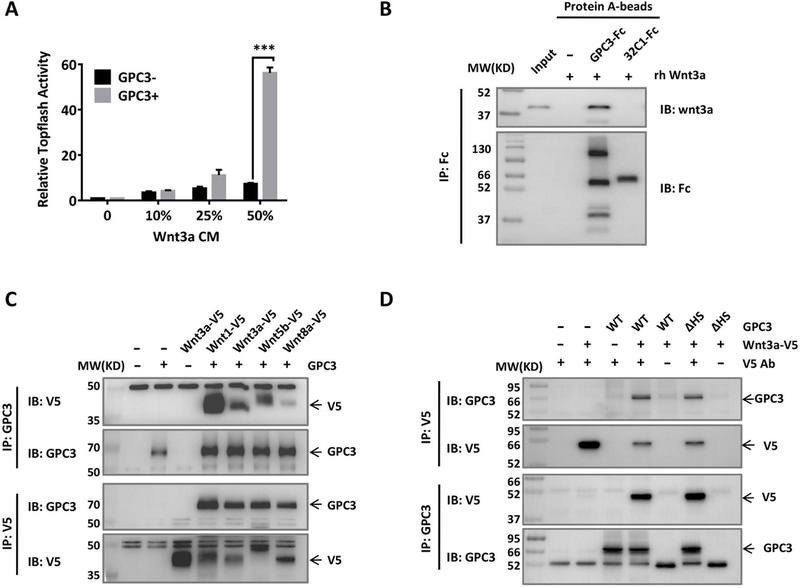
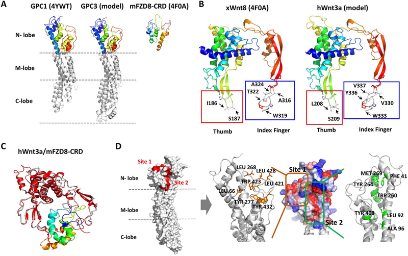
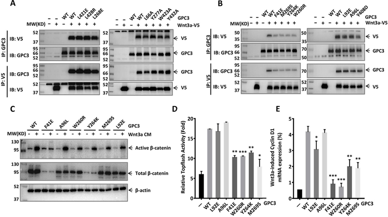
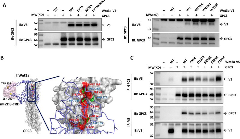
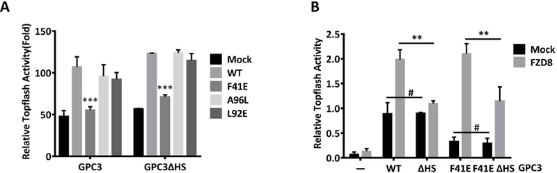
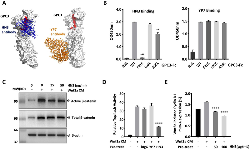
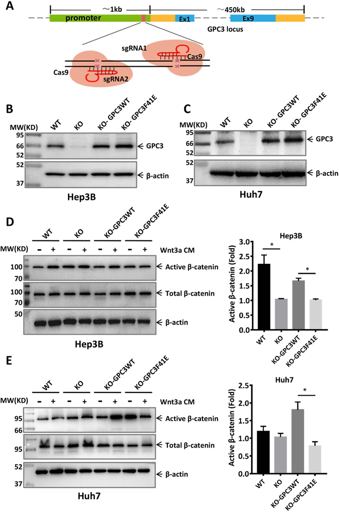
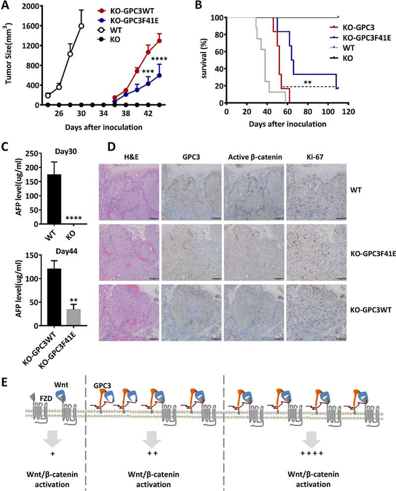
Wnt signaling is one of the key regulators of hepatocellular carcinoma (HCC) tumor progression. In addition to the classical receptor frizzled (FZD), various coreceptors including heparan sulfate proteoglycans (HSPGs) are involved in Wnt activation. Glypican-3 (GPC3) is an HSPG that is overexpressed in HCC and functions as a Wnt coreceptor that modulates HCC cell proliferation. These features make GPC3 an attractive target for liver cancer therapy. However, the precise interaction of GPC3 and Wnt and how GPC3, Wnt, and FZD cooperate with each other are poorly understood. In this study, we established a structural model of GPC3 containing a putative FZD-like cysteine-rich domain at its N-terminal lobe. We found that F41 and its surrounding residues in GPC3 formed a Wnt-binding groove that interacted with the middle region located between the lipid thumb domain and the index finger domain of Wnt3a. Mutating residues in this groove significantly inhibited Wnt3a binding, β-catenin activation, and the transcriptional activation of Wnt-dependent genes. In contrast with the heparan sulfate chains, the Wnt-binding groove that we identified in the protein core of GPC3 seemed to promote Wnt signaling in conditions when FZD was not abundant. Specifically, blocking this domain using an antibody inhibited Wnt activation. In HCC cells, mutating residue F41 on GPC3 inhibited activation of β-catenin in vitro and reduced xenograft tumor growth in nude mice compared with cells expressing wild-type GPC3. Conclusion: Our investigation demonstrates a detailed interaction of GPC3 and Wnt3a, reveals the precise mechanism of GPC3 acting as a Wnt coreceptor, and provides a potential target site on GPC3 for Wnt blocking and HCC therapy.


Conflict of interest statement

Conflict of interest:

The authors declare that they have no conflicts of interest with the contents of this article.

Figures

Fig. 1
Fig. 1. GPC3 interacted with Wnt and promoted Wnt activation.
A. Topflash assay to evaluate the effect of GPC3 on Wnt activation. Different amounts of Wnt3a CM were added to HEK293 Topflash cells transfected with or without GPC3. Luciferase activity was measured 24 h later. The data represent the mean ± SD (***P<0.001). B. Pull-down assays to detect the interaction of GPC3-Fc and recombinant human Wnt3a (rhWnt3a) in vitro. 32C1-Fc was used as an Fc control protein. The immunoprecipitated complex was detected with an anti-Wnt3a antibody or an anti-Fc antibody. C. Co-IP assay to detect the interactions of GPC3 with different Wnt ligands. HEK293T cells were transfected with various V5-tagged Wnt and full-length GPC3. Lysates from the cells were used for immunoprecipitation with anti-V5 or anti-GPC3 antibodies. D. Co-IP assay to detect the effect of HS chains on the interaction of GPC3 and Wnt3a. Wild-type GPC3 or GPC3ΔHS was co-transfected with V5-tagged Wnt3a. Lysates from the cells were used for immunoprecipitation with anti-V5 and anti-GPC3 antibodies.
Fig. 2
Fig. 2. Prediction of the potential Wnt-binding site on GPC3.
A. Comparison of GPC1 (PDB: 4YWT), GPC3 (model) and mFZD8-CRD (PDB: 4F0A). The GPC3 modeled structure was predicted by SWISS-MODEL. N-lobe: amino-terminal lobe; M-lobe: middle lobe; C-lobe: carboxy-terminal lobe. B. Modeled human Wnt3a structure. Left: X. laevis Wnt8 (PDB: 4F0A); right: modeled hWnt3a (predicted by SWISS-MODEL). The thumb sites were labeled with red squares, and the index finger sites were labeled with blue squares. C. The superimposed model for hWnt3a/mFZD8-CRD. The gray ribbon was the xWnt8 crystal structure, with lipid shown as red sticks. The red cartoon was the modeled hWnt3a, which was superimposed on xWnt8 in the crystal structure of xWnt8/mFZD8-CRD. mFZD8-CRD was shown in a rainbow-colored schematic. D. Two predicted regions with exposed hydrophobic surface in modeled GPC3. Left: Position of the two predicted hydrophobic regions (red) on the N-lobe of modeled GPC3. Right: A close-up view of the GPC3 N-lobe model. The surface was colored to indicate the hydrophobicity of each amino acid (red, most hydrophobic; blue, most hydrophilic). Site 1 (orange square) indicated the hydrophobic patch; site 2 (green square) indicated the region of the hydrophobic groove. Residues selected for mutation were labeled in orange and green, respectively (right).
Fig. 3.
Fig. 3.. Identification of the Wnt-binding area of GPC3.
Wild-type or mutant GPC3 were co-transfected with V5-tagged Wnt3a in HEK293T cells. Twenty-four hours later, the cells were lysed and incubated with GPC3 antibody or V5 antibody to detect the interaction of GPC3 and Wnt3a by Co-IP assay. A. Co-IP assay to detect the effect of site 1 on the interaction of GPC3 and Wnt3a. B. Co-IP assay to detect the effect of site 2 on the interaction of GPC3 and Wnt3a. C. HEK293 Topflash cells were transfected with indicated mutant GPC3. Twenty-four hours after the transfection, the cells were starved for 12 h and then stimulated with 50% Wnt3a CM. Twelve hours later, the expression levels of active β-catenin (C), Topflash reporter activity (D), and mRNA expression level of Cyclin D1 (E) were measured. The results are represented at least three independent experiments. The data represent the mean ± SD (*P<0.05, **P<0.01 and ***P<0.001).
Fig. 4.
Fig. 4.. Identification of the GPC3-binding area of Wnt3a.
A. Co-IP assay to detect the effect of two finger-like domains of Wnt3a on GPC3 interaction. V5-tagged Wnt3a with a point mutation in the lipid thumb domain (S209) or in the index finger domain (W333) was co-transfected with GPC3 in HEK293T cells. Residue C77 is another palmitoylated modification site of Wnt3a(36). Twenty-four hours later, the cells were lysed and incubated with GPC3 antibody or V5 antibody to detect the interaction of GPC3 and Wnt3a. B. The predicted model of mFZD8-CRD/hWnt3a/GPC3 complex. GPC3 and mFZD8-CRD were shown in surface representation (mFZD8-CRD in pink and GPC3 in gray), and hWnt3a was shown with the purple cartoon/stick representation. The residues in Wnt3a essential for GPC3 binding were labeled in blue. The residues in the region of the hydrophobic groove (site 2) for Wnt binding were labeled in red. C. V5-tagged Wnt3a with the point mutations shown in B was co-transfected with GPC3 in HEK293T cells. The interaction of GPC3 and Wnt3a was detected by Co-IP with the method described in A.
Fig. 5.
Fig. 5.. The influence of HS chains of GPC3 on F41-mediated Wnt activation.
A. Topflash reporter assay to detect the effect of the mutation F41E with or without HS chains on Wnt activation. HEK293 Topflash cells were transfected with wild-type GPC3 or GPC3ΔHS carrying the indicated point mutation. Twenty-four hours after the transfection, the cells were starved for 12 h and luciferase activity was measure after stimulation with 50% Wnt3a CM for 12h. The data represent the mean ± SD (***P<0.001). B. Topflash assay to compare the effect of HS chain-mediated and F41-mediated Wnt activation with or without mFZD8. HEK293 Topflash cells were co-transfected with the indicated mutant GPC3 and mFZD8. The cells were then treated as described in A. The data represent the mean ± SD (**P<0.01, #P>0.05).
Fig. 6.
Fig. 6.. Antibody HN3 recognized the Wnt-binding area of GPC3 and blocked Wnt activation.
A. Putative binding of HN3 and YP7 on modeled GPC3. The Wnt-binding area (site 2) was labeled in red. Left: HN3 (blue stick) was docked on the modeled GPC3 by setting its three CDRs as the interactive interface. Right: YP7 (orange stick) was docked on the modeled full-length GPC3 (by Phyre2) by setting its six CDRs as the interface. The sequence corresponding to the peptide used to immunize mice for generating YP7 was labeled in grey. B. ELISA to detect HN3 and YP7 binding activity on wild type and site 2 mutant GPC3. Purified wild-type GPC3 or the indicated GPC3 mutant proteins were coated on ELISA plates. After blocking, HN3 or YP7 were added to detect the binding activity at OD450 nm. The results are represented at least three independent experiments. The data represent the mean ± SD (**P<0.01 and ***P<0.001). C. Western blot to detect the blocking effect of HN3 on Wnt3a-induced β-catenin activation. HEK293 Topflash-GPC3 stable cells were starved overnight and pre-treated with HN3 for 30 minutes. Then 50% Wnt3a CM were added. Six hours later, cells were harvested to detect the expression of active β-catenin (C), Topflash reporter activity (D) and mRNA expression of Cyclin D1 (E). The results are represented at least three independent experiments. The data represent the mean ± SD (**P<0.01).
Fig. 7.
Fig. 7.. GPC3F41E caused impaired Wnt activation in HCC cells.
A. Schematic for constructing GPC3 knockout cell lines using CRISPR-Cas9. Two sgRNAs were designed to target the promoter of the GPC3 gene. B. Re-expression of GPC3WT and GPC3F41E in GPC3 knockout Hep3B cells (clone 12). C. Re-expression of GPC3WT and GPC3F41E in GPC3 knockout Huh-7 cells (clone 15). WT: wild type, KO: knockout. Cells constructed in B and C were starved overnight and then treated with or without 50% Wnt3a CM for 12 h. Western blot to show the expression of active β-catenin in D and E, respectively. The statistical analyses were shown as fold change by density calculating from three independent experiments. The data represent the mean ± SD (*P<0.05).
Fig. 8.
Fig. 8.. GPC3F41E inhibited HCC tumor growth in mice.
BALB/c nu/nu mice were subcutaneously inoculated with 5×10⁶ WT, KO, KO-GPC3WT, and KO-GPC3F41E Hep3B cells. A. Tumor growth curve. WT n=11; KO n=9; KO-GPC3WT n=9; KO-GPC3F41E n=8. The data represent the mean ± SE (***P<0.001 and ****P<0.0001). B. Survival curve. WT n=8; KO n=9; KO-GPC3WT n=6; KO-GPC3F41E n=6 (**P<0.01 and ****P<0.0001). C. AFP level of mice at day 30 (WT n=11; KO n=9) and day 44 (KO-GPC3WT n=7; KO-GPC3F41E n=6). The data represent the mean ± SE (**P<0.01 and ****P<0.0001). D. Hematoxylin and eosin staining and immunohistochemistry staining for GPC3, active β-catenin and Ki-67 in the tumors from A. Scale bar, 200 μm. E. The working model of GPC3 regulating Wnt activation in HCC cells.

Similar articles

Cited by

References

    1. Global Burden of Disease Cancer C, Fitzmaurice C, Allen C, Barber RM, Barregard L, Bhutta ZA, Brenner H, et al. Global, Regional, and National Cancer Incidence, Mortality, Years of Life Lost, Years Lived With Disability, and Disability-Adjusted Life-years for 32 Cancer Groups, 1990 to 2015: A Systematic Analysis for the Global Burden of Disease Study. JAMA Oncol 2017;3:524–548. - PMC - PubMed
    1. Forner A, Llovet JM, Bruix J. Hepatocellular carcinoma. Lancet 2012;379:1245–1255. - PubMed
    1. Torre LA, Bray F, Siegel RL, Ferlay J, Lortet-Tieulent J, Jemal A. Global cancer statistics, 2012. CA Cancer J Clin 2015;65:87–108. - PubMed
    1. Filmus J, Capurro M, Rast J. Glypicans. Genome Biol 2008;9:224. - PMC - PubMed
    1. Ho M, Kim H. Glypican-3: a new target for cancer immunotherapy. Eur J Cancer 2011;47:333–338. - PMC - PubMed

Publication types

MeSH terms