Review
. 2019 May;285(5):479-490.
doi: 10.1111/joim.12910. Epub 2019 Apr 22.

Mucus and mucins in diseases of the intestinal and respiratory tracts

Affiliations
Review

Mucus and mucins in diseases of the intestinal and respiratory tracts

G C Hansson. J Intern Med. 2019 May.

Abstract

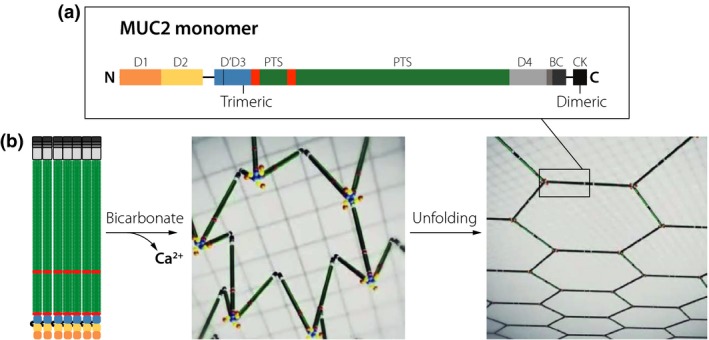
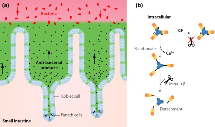
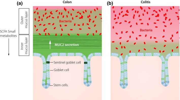
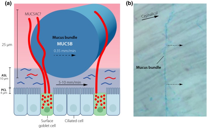
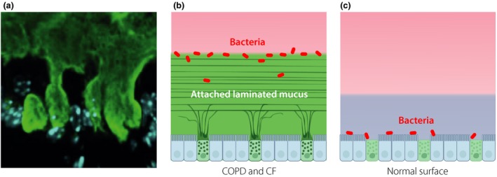
This review describes the organization and importance of mucus in the intestine and lungs in relation to the diseases cystic fibrosis (CF), ulcerative colitis and chronic obstructive pulmonary disease (COPD). The inner surfaces of the body are protected by mucus built around polymeric glycoproteins called mucins. In the disease CF, the small intestinal mucus is in contrast the normal attached to the epithelium, explaining the intestinal problems at this disease. The inner of the two mucus layers of colon is normally impenetrable to bacteria, keeping the commensals away from and protecting the epithelium. This impenetrable property is dependent on the bacterial composition and the host diet, observations that can explain the increased incidence of inflammatory bowel diseases in the western world as bacteria reach the epithelial cells in active ulcerative colitis. The respiratory tract is normally cleared by thick mucus bundles that moved by the cilia sweep the epithelial surface. In CF, the bundles are nonmoving already at birth. Cholinergic stimulations stop the bundle movement explaining some of the beneficial effect of anticholinergic treatment in COPD. In this disease as well as in more developed CF, an attached mucus layer is formed. This mucus has features similar to the protective inner colon mucus and is by this able to separate bacteria from the epithelial surface. When formed in healthy individuals this mucus can be coughed up, but in chronically diseased lungs, bacteria colonizing the mucus will remain in the lungs and the resulting inflammation contribute to the destruction of the lungs.

Keywords: COPD; cystic fibrosis; mucin; mucus; ulcerative colitis.


Conflict of interest statement

No conflict of interest was declared.

Figures

Figure 1
Figure 1
The MUC2 mucin structure, intracellular packing and unfolding upon secretion. (a) Domain organization of the 5130 amino acids of the MUC2 mucin, N, N‐terminal; C, C‐terminal; D. von Willebrand D domain; B, von Willebrand B domain; PTS, proline, threonine, serine sequence that after glycosylation become a mucin domain; CK, cysteine knot domain. (b) MUC2 mucin drawn to scale and packed in the secretory granule. After secretion, bicarbonate helps to remove calcium from MUC2 by increasing the pH and binding calcium allowing the mucin polymer to unfold into a flat net‐like structure that organizes the inner colon mucus layer.
Figure 2
Figure 2
The mucus system of the small intestine. (a) All spaces between the small intestinal villi and over the villi tips are filled out by mucus generating a diffusion barrier that do not exclude bacteria by its pore size. Paneth cells in the crypt bottom secrete antibacterial peptides and proteins that together with the mucus keep the bacteria away from the epithelial cells. (b) The mucus of the small intestine is normally detached from its attachment by the meprin β protease cleaving off the N‐terminus of MUC2. Sufficient amounts of bicarbonate are required to unfold the MUC2 N‐termini and by this expose the meprin β cleavage site. In cystic fibrosis (CF), this unfolding is insufficient and renders the small intestine attached. Colours of the MUC2 N‐terminal domains as in Fig. 1.
Figure 3
Figure 3
The mucus of the colon is two layered with an inner attached stratified mucus layer and a nonattached outer mucus layer. (a) The inner layer has pore sizes not allowing bacteria to enter, whereas the outer layer is expanded and generating the habitat for the commensal bacteria. Every crypt opening is guarded by a Sentinel goblet cell that senses when increased bacteria come in close contact and after activation coordinate compound exocytosis of a ring of goblet cells to generate a mucus plume moving bacteria away. The bacteria in the outer mucus layer generate short fatty acids (SCFA) and other metabolites that affect the epithelium. (b) In active ulcerative colitis, bacteria are reaching the epithelium due to failure of the inner mucus layer and loss of Sentinel goblet cells.
Figure 4
Figure 4
The mucus protective system of normal upper respiratory tract of higher animals. (a) Submucosal glands generate mucus bundles by >1000 MUC5B mucin linear polymers. The bundles are moved by the cilia and the air surface liquid (ASL). The mucus bundles are coated by the MUC5AC mucin from the surface goblet cells. (b) Mucus bundles stained with Alcian blue are moved cephalically over the tracheobronchial surface.
Figure 5
Figure 5
In chronic lung diseases like chronic obstructive pulmonary disease (COPD) and cystic fibrosis (CF), a mucus layer is attached to the surface epithelium. (a) Stained mucus anchored to surface goblet cells. (b) Schematic picture showing an attached laminated mucus layer where bacteria are trapped on and in the mucus. (c) On normal lungs surface, bacteria can come in direct contact with the epithelial cells. Blue layer is air surface liquid.

Similar articles

Cited by

References

    1. Johansson MEV, Phillipson M, Petersson J, Holm L, Velcich A, Hansson GC. The inner of the two Muc2 mucin dependent mucus layers in colon is devoid of bacteria. Proc Natl Acad Sci USA 2008; 105: 15064–9. - PMC - PubMed
    1. Johansson MEV, Thomsson KA, Hansson GC. Proteomic analyses of the two mucus layers of the colon barrier reveal that their main component, the Muc2 mucin, is strongly bound to the Fcgbp protein. J Proteome Res 2009; 8: 3549–57. - PubMed
    1. Pigny P, Guyonnetduperat V, Hill AS et al Human mucin genes assigned to 11p15.5: identification and organization of a cluster of genes. Genomics 1996; 38: 340–52. - PubMed
    1. Lang T, Hansson GC, Samuelsson T. Gel‐forming mucins appeared early in metazoan evolution. Proc Natl Acad Sci USA 2007; 104: 16209–14. - PMC - PubMed
    1. Lang T, Klasson S, Larsson E, Johansson MEV, Hansson GC, Samuelsson T. Searching the evolutionary origin of epithelial mucus protein components‐mucins and FCGBP. Mol Biol Evol 2016; 33: 1921–36. - PMC - PubMed

Publication types

MeSH terms