

Role and prospects of regenerative biomaterials in the repair of spinal cord injury
- PMID: 30964053
- PMCID: PMC6524500
- DOI: 10.4103/1673-5374.253512
Role and prospects of regenerative biomaterials in the repair of spinal cord injury
Abstract
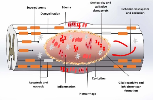
Axonal junction defects and an inhibitory environment after spinal cord injury seriously hinder the regeneration of damaged tissues and neuronal functions. At the site of spinal cord injury, regenerative biomaterials can fill cavities, deliver curative drugs, and provide adsorption sites for transplanted or host cells. Some regenerative biomaterials can also inhibit apoptosis, inflammation and glial scar formation, or further promote neurogenesis, axonal growth and angiogenesis. This review summarized a variety of biomaterial scaffolds made of natural, synthetic, and combined materials applied to spinal cord injury repair. Although these biomaterial scaffolds have shown a certain therapeutic effect in spinal cord injury repair, there are still many problems to be resolved, such as product standards and material safety and effectiveness.
Keywords: combination; functional recovery; microenvironment; nerve regeneration; neural regeneration; regeneration; regenerative biomaterials; repair strategy; scaffolds; spinal cord injury; tissue engineering; transplantation.
Conflict of interest statement
None
Figures


References
-
- Agbay A, Edgar JM, Robinson M, Styan T, Wilson K, Schroll J, Ko J, Mohtaram NK, Jun BG, Willerth SM. Biomaterial strategies for delivering stem cells as a treatment for spinal cord injury. Cells Tissues Organs. 2016;202:42. - PubMed
-
- Alhosseini SN, Moztarzadeh F, Mozafari M, Asgari S, Dodel M, Samadikuchaksaraei A, Kargozar S, Jalali N. Synthesis and characterization of electrospun polyvinyl alcohol nanofibrous scaffolds modified by blending with chitosan for neural tissue engineering. Int J Nanomedicine. 2012;7:25–34. - PMC - PubMed
-
- Amr SM, Ashraf G, Koptan WT, Galal AA, Dina Sabry AF, Rashed LA, Atta HM, Abdel-Aziz MT. Bridging defects in chronic spinal cord injury using peripheral nerve grafts combined with a chitosan-laminin scaffold and enhancing regeneration through them by co-transplantation with bone-marrow-derived mesenchymal stem cells: case series of 14 patients. J Spinal Cord Med. 2014;37:54–71. - PMC - PubMed
-
- Ando K, Imagama S, Ito Z, Kobayashi K, Hida T, Nakashima H, Ito K, Tsushima M, Ishikawa Y, Matsumoto A, Nishida K, Nishida Y, Ishiguro N. Self-assembling peptide reduces glial scarring, attenuates posttraumatic inflammation, and promotes neurite outgrowth of spinal motor neurons. Spine. 2016;41:E1201–E1207. - PubMed
-
- Bakshi A, Fisher O, Dagci T, Himes BT, Fischer I, Lowman A. Mechanically engineered hydrogel scaffolds for axonal growth and angiogenesis after transplantation in spinal cord injury. J Neurosurg Spine. 2004;1:322–329. - PubMed
Publication types
LinkOut - more resources
Full Text Sources
