. 2019 Aug;14(8):1455-1461.
doi: 10.4103/1673-5374.253531.

Variation in expression of small ubiquitin-like modifiers in injured sciatic nerve of mice

Affiliations

Variation in expression of small ubiquitin-like modifiers in injured sciatic nerve of mice

Dian-Ying Zhang et al. Neural Regen Res. 2019 Aug.

Abstract

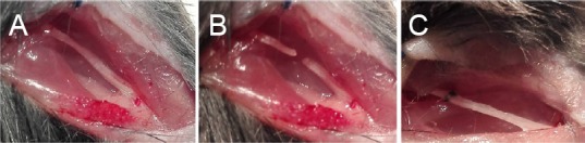
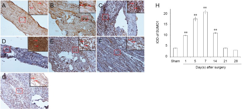
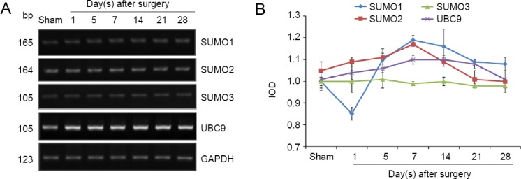
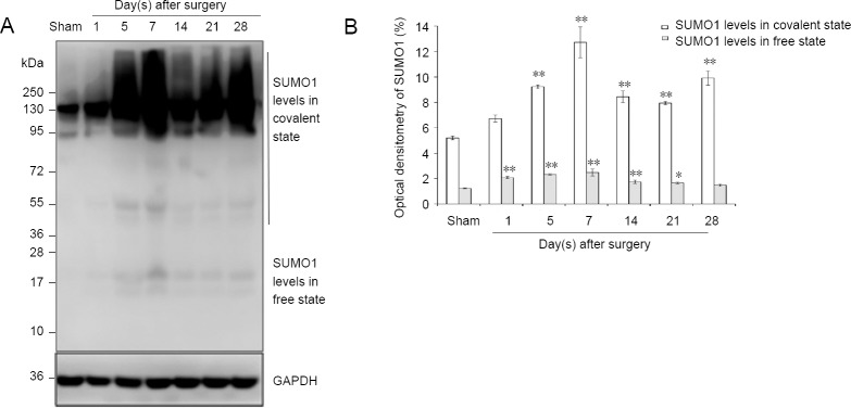
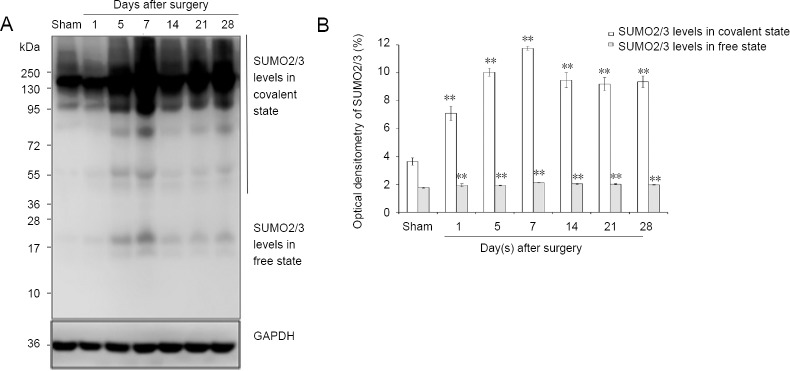
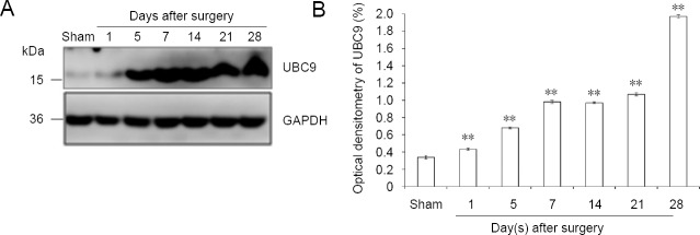
Small ubiquitin-like modifiers (SUMOs) have been shown to regulate axonal regeneration, signal transduction, neuronal migration, and myelination, by covalently and reversibly attaching to the protein substrates during neuronal cell growth, development, and differentiation. It has not been reported whether SUMOs play a role in peripheral nerve injury and regeneration. To investigate any association between SUMOylation and potential neuroprotective effects during peripheral nerve injury and regeneration, C57/BL mice were randomly divided into sham and experimental groups. The sciatic nerve was exposed only in the sham group. The experimental group underwent neurotomy and epineurial neurorrhaphy. Real-time quantitative polymerase chain reaction and western blot assay results revealed different mRNA and protein expression levels of SUMO1, SUMO2, SUMO3 and UBC9 in sciatic nerve tissue (containing both 5 mm of proximal and distal stumps at the injury site) at various time points after injury. Compared with the sham group, protein levels of SUMO1 and SUMO2/3 increased in both their covalent and free states after sciatic nerve injury in the experimental group, especially in the covalent state. UBC9 protein levels showed similar changes to those of SUMO1 and SUMO2/3 in the covalent states. Immunohistochemical staining demonstrated that SUMO1 and SUMO2/3 immunopositivities were higher in the experimental group than in the sham group. Our results verified that during the repair of sciatic nerve injury, the mRNA and protein expression of SUMO1, SUMO2, SUMO3 and UBC9 in injured nerve tissues changed in varying patterns and there were clear changes in the expression of SUMO-related proteins. These findings reveal that SUMOs possibly play an important role in the repair of peripheral nerve injury. All animal protocols were approved by the Institutional Animal Care and Use Committee of Tianjin Fifth Central Hospital, China (approval No. TJWZXLL2018041) on November 8, 2018.

Keywords: SUMO1; SUMO2/3; SUMOylation; UBC9; epineurial neurorrhaphy; nerve regeneration; neural regeneration; peripheral nerve injury; sciatic nerve.


Conflict of interest statement

None

Figures

Figure 1
Figure 1
Suture model of sciatic nerve. (A) Sciatic nerve exposure; (B) sciatic nerve transection at the sciatic notch; (C) epineurial neurorrhaphy of the transected sciatic nerve.
Figure 2
Figure 2
Immunohistochemical staining for SUMO1 in sciatic nerves. (A) SUMO1 immunopositive staining (arrows) in sciatic nerve of the sham group (1 cm sciatic nerves around the sciatic notch); (B–G) SUMO1 immunopositive staining (arrows) in the sciatic nerve (containing both 5 mm proximal and distal stumps at the injury site) at 1, 5, 7, 14, 21, and 28 days after sciatic nerve injury in the experimental group, respectively. SUMO1 immunopositive staining was observed in A–G, especially in the sciatic nerve suture point (the boxed areas) of Figure B–E. Using optical microscope, original magnification, 20×; insets are higher magnification of the boxed areas, original magnification, 100×. (H) Quantitative analysis of SUMO1 immunopositivity in each group. **P < 0.01, vs. sham group (mean ± SD, n = 4, one-way analysis of variance followed by Tukey’s honestly significant difference post hoc test). IOD: Integrated optical density; SUMO: small ubiquitin-like modifier.
Figure 3
Figure 3
Immunohistochemical staining for SUMO2/3 in the sciatic nerve. (A) SUMO2/3 immunopositive staining (arrows) in the sciatic nerve of sham group (1 cm sciatic nerves around the sciatic notch); (B–G) SUMO2/3 immunopositive staining (arrows) in the sciatic nerve (containing both 5 mm proximal and distal stumps at the injury site) at 1, 5, 7, 14, 21 and 28 days after sciatic nerve injury in the experimental group, respectively. SUMO2/3 immunopositive staining was observed in A–G, especially at the sciatic nerve suture point (the boxed areas) of B–G. Using optical microscope, original magnification, 20×; insets are higher magnification of the boxed areas, original magnification, 100×. (H) Quantitative analysis of SUMO2/3 immunopositivity in each group. **P < 0.01, vs. sham group (mean ± SD, n = 4, one-way analysis of variance followed by Tukey’s honestly significant difference post hoc test). IOD: Integrated optical density; SUMO: small ubiquitin-like modifier.
Figure 4
Figure 4
SUMO1, SUMO2, SUMO3, and UBC9 mRNA expression levels after sciatic nerve injury. (A) Real-time quantitative polymerase chain reaction analyses for SUMO1, SUMO2, SUMO3, and UBC9 of the sciatic nerve. (B) Quantification of expression of SUMO1, SUMO2, SUMO3, and UBC9 in each group: GAPDH was used as an internal control. UBC9: Ubiquitin-conjugating enzyme 9; GAPDH: glyceraldehyde-3-phosphate dehydrogenase; SUMO: small ubiquitin-like modifier.
Figure 5
Figure 5
SUMO1 protein expression in sciatic nerve. (A) Detection of SUMO1 levels in covalent and free states by western blot assay. (B) Quantification of expression of SUMO1 in each group, GAPDH was used as an internal control. *P < 0.05, **P < 0.01, vs. sham group (mean ± SD, n = 4, one-way analysis of variance followed by Tukey’s honestly significant difference post hoc test). SUMO: Small ubiquitin-like modifier; GAPDH: glyceraldehyde-3-phosphate dehydrogenase.
Figure 6
Figure 6
SUMO2/3 protein expression in sciatic nerve. (A) Detection of SUMO2/3 levels in covalent and free states by western blot assay. (B) Quantification of expression of SUMO2/3 in each group: GAPDH was used as an internal control. **P < 0.01, vs. sham group (mean ± SD, n = 4, one-way analysis of variance followed by Tukey’s honestly significant difference post hoc test). SUMO: Small ubiquitin-like modifier; GAPDH: glyceraldehyde-3-phosphate dehydrogenase; d: day(s).
Figure 7
Figure 7
UBC9 protein expression in sciatic nerve. (A) Detection of UBC9 levels by western blot assay. (B) Quantification of expression of UBC9 in each group: GAPDH was used as an internal control. **P < 0.01, vs. sham group (mean ± SD, n = 4, one-way analysis of variance followed by Tukey’s honestly significant difference post hoc test). UBC9: Ubiquitin-conjugating enzyme; GAPDH: glyceraldehyde-3-phosphate dehydrogenase.

Similar articles

Cited by

References

    1. Anderson DB, Wilkinson KA, Henley JM. Protein SUMOylation in neuropathological conditions. Drug News Perspect. 2009;22:255–265. - PMC - PubMed
    1. Anderson DB, Zanella CA, Henley JM, Cimarosti H. Sumoylation: implications for neurodegenerative diseases. Adv Exp Med Biol. 2017;963:261–281. - PubMed
    1. Andreassi C, Crerar H, Riccio A. Post-transcriptional processing of mRNA in neurons: the vestiges of the RNA world drive transcriptome diversity. Front Mol Neurosci. 2018;11:304. - PMC - PubMed
    1. Bosse F, Kury P, Muller HW. Gene expression profiling and molecular aspects in peripheral nerve regeneration. Restor Neurol Neurosci. 2001;19:5–18. - PubMed
    1. Bosse F, Hasenpusch-Theil K, Kury P, Muller HW. Gene expression profiling reveals that peripheral nerve regeneration is a consequence of both novel injury-dependent and reactivated developmental processes. J Neurochem. 2006;96:1441–1457. - PubMed