

Mesoporous bioactive glass/ɛ-polycaprolactone scaffolds promote bone regeneration in osteoporotic sheep
- PMID: 30965142
- PMCID: PMC6726488
- DOI: 10.1016/j.actbio.2019.04.019
Mesoporous bioactive glass/ɛ-polycaprolactone scaffolds promote bone regeneration in osteoporotic sheep
Abstract
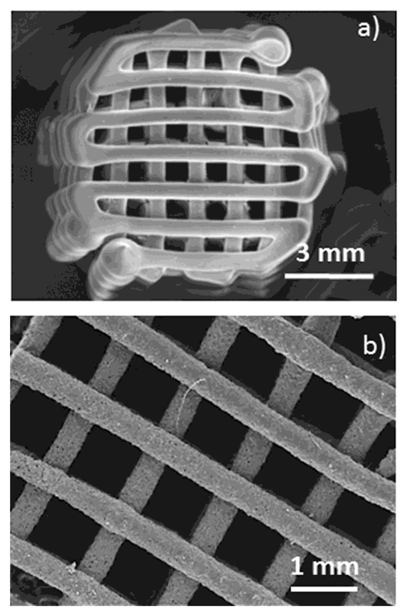
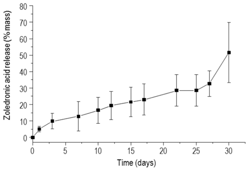
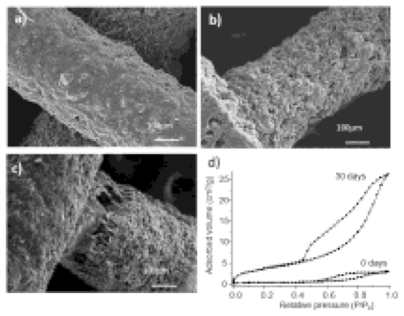
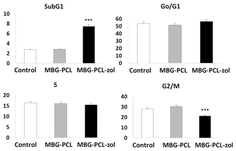
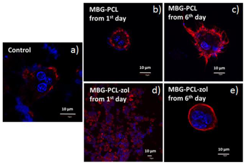
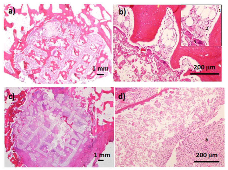
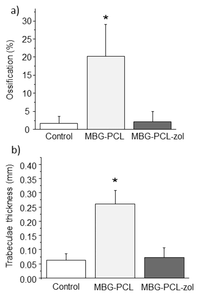
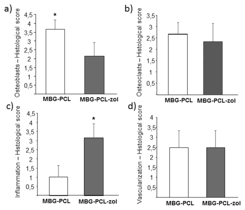
Macroporous scaffolds made of a SiO2-CaO-P2O5 mesoporous bioactive glass (MBG) and ɛ-polycaprolactone (PCL) have been prepared by robocasting. These scaffolds showed an excellent in vitro biocompatibility in contact with osteoblast like cells (Saos 2) and osteoclasts derived from RAW 264.7 macrophages. In vivo studies were carried out by implantation into cavitary defects drilled in osteoporotic sheep. The scaffolds evidenced excellent bone regeneration properties, promoting new bone formation at both the peripheral and the inner parts of the scaffolds, thick trabeculae, high vascularization and high presence of osteoblasts and osteoclasts. In order to evaluate the effects of the local release of an antiosteoporotic drug, 1% (%wt) of zoledronic acid was incorporated to the scaffolds. The scaffolds loaded with zoledronic acid induced apoptosis in Saos 2 cells, impeded osteoclast differentiation in a time dependent manner and inhibited bone healing, promoting an intense inflammatory response in osteoporotic sheep. STATEMENT OF SIGNIFICANCE: In addition to an increase in bone fragility and susceptibility to fracture, osteoporosis also hinders the clinical success of endosseous implants and grafting materials for the treatment of bone defects. For the first time, macroporous scaffolds made of mesoporous bioactive glass and ε-caprolactone have been evaluated in a sheep model that mimics the osteoporosis conditions in humans. These implants fostered bone regeneration, promoting new bone formation at both the peripheral and the inner parts of the scaffolds, showing thick trabeculae and a high vascularization degree. Our results indicate that macroporous structures containing highly bioactive mesoporous glasses could be excellent candidates for the regenerative treatment of bone defects in osteoporotic patients.
Keywords: Bone regeneration; Mesoporous bioactive glass; Osteoporosis; Scaffolds; Zoledronic acid.
Copyright © 2019 Acta Materialia Inc. Published by Elsevier Ltd. All rights reserved.
Figures
References
-
- Consensus development conference: Diagnosis, prophylaxis, and treatment of osteoporosis. Am J Med. 1993;94:646–650. - PubMed
-
- Ismail AA, Cooper C, Felsenberg D, Varlow J, Kanis JA, Silman AJ, O’Neill TW. Number and type of vertebral deformities: epidemiological characteristics and relation to back pain and height loss. European Vertebral Osteoporosis Study Group. Osteoporos Int. 1999;9:206–213. - PubMed
-
- Ettinger B, Black DM, Mitlak BH, Knickerbocker RK, Nickelsen T, Genant HK, Christiansen C, Delmas PD, Zanchetta JR, Stakkestad J, Glüer CC, et al. Reduction of vertebral fracture risk in postmenopausal women with osteoporosis treated with raloxifene: results from a 3-year randomized clinical trial. Multiple Outcomes of Raloxifene Evaluation (MORE) Investigators. JAMA. 1999;282:637–645. - PubMed
-
- Fini M, Giavaresi G, Torricelli P, Krajewski A, Ravaglioli A, Belmonte MM, Biagini G, Giardino R. Biocompatibility and osseointegration in osteoporotic bone. A preliminary in vitro and in vivo study. J Bone Joint Surg [Br] 2001;83-B:139–143. - PubMed
-
- Marco F, Milena F, Gianluca G, Vittoria O. Peri-implant osteogenesis in health and osteoporosis. Micron. 2005;36:630–644. - PubMed
Publication types
MeSH terms
Substances
Grants and funding
LinkOut - more resources
Full Text Sources
Medical
Research Materials
