

Biofunctional Nanofibrous Substrate for Local TNF-Capturing as a Strategy to Control Inflammation in Arthritic Joints
- PMID: 30965588
- PMCID: PMC6523323
- DOI: 10.3390/nano9040567
Biofunctional Nanofibrous Substrate for Local TNF-Capturing as a Strategy to Control Inflammation in Arthritic Joints
Abstract
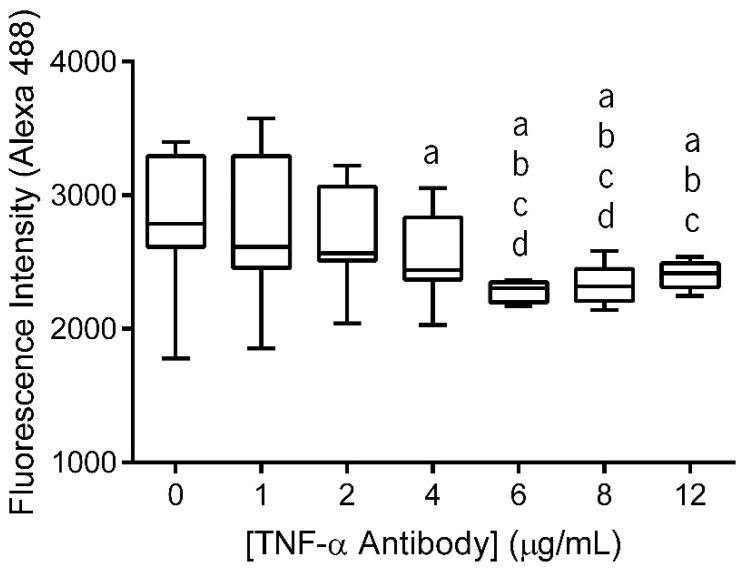
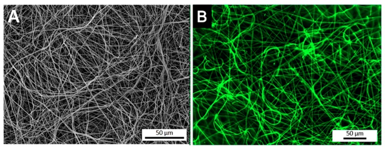
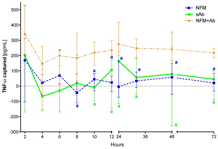
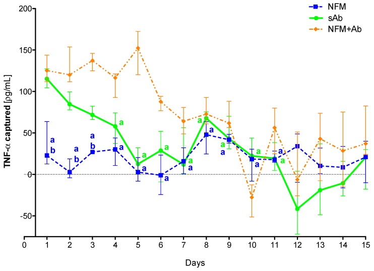
Rheumatoid arthritis (RA) is an autoimmune disease that affects the synovial cavity of joints, and its pathogenesis is associated with an increased expression of pro-inflammatory cytokines, namely tumour necrosis factor-alpha (TNF-α). It has been clinically shown to have an adequate response to systemic administration of TNF-α inhibitors, although with many shortcomings. To overcome such limitations, the immobilization of a TNF-α antibody on a nanofibrous substrate to promote a localized action is herein proposed. By using this approach, the antibody has its maximum therapeutic efficacy and a prolonged therapeutic benefit, avoiding the systemic side-effects associated with conventional biological agents' therapies. To technically achieve such a purpose, the surface of electrospun nanofibers is initially activated and functionalized, allowing TNF-α antibody immobilization at a maximum concentration of 6 µg/mL. Experimental results evidence that the biofunctionalized nanofibrous substrate is effective in achieving a sustained capture of soluble TNF-α over time. Moreover, cell biology assays demonstrate that this system has no deleterious effect over human articular chondrocytes metabolism and activity. Therefore, the developed TNF-capturing system may represent a potential therapeutic approach for the local management of severely affected joints.
Keywords: TNF-α capture; antibody immobilization; electrospun nanofibers; human articular chondrocytes; rheumatoid arthritis.
Conflict of interest statement
The authors declare no conflict of interest. The funders had no role in the design of the study; in the collection, analyses, or interpretation of data; in the writing of the manuscript, or in the decision to publish the results.
Figures
Similar articles
-
Mast cell activation and its relation to proinflammatory cytokine production in the rheumatoid lesion.Arthritis Res. 2000;2(1):65-74. doi: 10.1186/ar70. Arthritis Res. 2000. PMID: 11219391 Free PMC article.
-
Local versus systemic anti-tumour necrosis factor-α effects of adalimumab in rheumatoid arthritis: pharmacokinetic modelling analysis of interaction between a soluble target and a drug.Clin Pharmacokinet. 2012 Jul 1;51(7):443-55. doi: 10.2165/11599970-000000000-00000. Clin Pharmacokinet. 2012. PMID: 22540283
-
Periodontal Disease as a Risk Factor for Rheumatoid Arthritis: A Systematic Review.JBI Libr Syst Rev. 2012;10(42 Suppl):1-12. doi: 10.11124/jbisrir-2012-288. JBI Libr Syst Rev. 2012. PMID: 27820156
-
Anti-TNF alpha therapy of rheumatoid arthritis: what have we learned?Annu Rev Immunol. 2001;19:163-96. doi: 10.1146/annurev.immunol.19.1.163. Annu Rev Immunol. 2001. PMID: 11244034 Review.
-
Monoclonal anti-TNF alpha antibody as a probe of pathogenesis and therapy of rheumatoid disease.Immunol Rev. 1995 Apr;144:195-223. doi: 10.1111/j.1600-065x.1995.tb00070.x. Immunol Rev. 1995. PMID: 7590814 Review.
Cited by
-
Sustained Local Delivery of Diclofenac from Three-Dimensional Ultrafine Fibrous Protein Scaffolds with Ultrahigh Drug Loading Capacity.Nanomaterials (Basel). 2019 Jun 26;9(7):918. doi: 10.3390/nano9070918. Nanomaterials (Basel). 2019. PMID: 31247985 Free PMC article.
-
Decellularized Human Chorion Membrane as a Novel Biomaterial for Tissue Regeneration.Biomolecules. 2020 Aug 20;10(9):1208. doi: 10.3390/biom10091208. Biomolecules. 2020. PMID: 32825287 Free PMC article.
-
Stimulation of Neurite Outgrowth Using Autologous NGF Bound at the Surface of a Fibrous Substrate.Biomolecules. 2021 Dec 24;12(1):25. doi: 10.3390/biom12010025. Biomolecules. 2021. PMID: 35053173 Free PMC article.
-
Transdermal delivery of inflammatory factors regulated drugs for rheumatoid arthritis.Drug Deliv. 2022 Dec;29(1):1934-1950. doi: 10.1080/10717544.2022.2089295. Drug Deliv. 2022. PMID: 35757855 Free PMC article.
-
Translation Research in Therapeutic Approaches from Conventional to Novel Nano-therapeutics for Rheumatoid Arthritis Treatment.Curr Rheumatol Rev. 2025;21(1):37-53. doi: 10.2174/0115733971288433240408062359. Curr Rheumatol Rev. 2025. PMID: 38629371 Review.
References
Grants and funding
LinkOut - more resources
Full Text Sources
Other Literature Sources
