. 2017 Oct 4;9(10):487.
doi: 10.3390/polym9100487.

Incorporation of Calcium Containing Mesoporous (MCM-41-Type) Particles in Electrospun PCL Fibers by Using Benign Solvents

Affiliations

Incorporation of Calcium Containing Mesoporous (MCM-41-Type) Particles in Electrospun PCL Fibers by Using Benign Solvents

Liliana Liverani et al. Polymers (Basel). .

Abstract

The electrospinning technique is a versatile method for the production of fibrous scaffolds able to resemble the morphology of the native extra cellular matrix. In the present paper, electrospinning is used to fabricate novel SiO2 particles (type MCM-41) containing poly(epsilon-caprolactone) (PCL) fibers. The main aims of the present work are both the optimization of the particle synthesis and the fabrication of composite fibers, obtained using benign solvents, suitable as drug delivery systems and scaffolds for soft tissue engineering applications. The optimized synthesis and characterization of calcium-containing MCM-41 particles are reported. Homogeneous bead-free composite electrospun mats were obtained by using acetic acid and formic acid as solvents; neat PCL electrospun mats were used as control. Initially, an optimization of the electrospinning environmental parameters, like relative humidity, was performed. The obtained composite nanofibers were characterized from the morphological, chemical and mechanical points of view, the acellular bioactivity of the composite nanofibers was also investigated. Positive results were obtained in terms of mesoporous particle incorporation in the fibers and no significant differences in terms of average fiber diameter were detected between the neat and composite electrospun fibers. Even if the Ca-containing MCM-41 particles are bioactive, this property is not preserved in the composite fibers. In fact, during the bioactivity assessment, the particles were released confirming the potential application of the composite fibers as a drug delivery system. Preliminary in vitro tests with bone marrow stromal cells were performed to investigate cell adhesion on the fabricated composite mats, the positive obtained results confirmed the suitability of the composite fibers as scaffolds for soft tissue engineering.

Keywords: benign solvents; composites; electrospinning; mesoporous silica calcium containing MCM-41; nanofibers; poly(epsilon-caprolactone).


Conflict of interest statement

The authors declare no conflict of interest.

Figures

Figure 1
Figure 1
SEM micrographs of calcium-containing MCM-41 (Ca_MCM-41) particles at low magnification. (a) higher magnification; (b) show the homogeneity of particles size and shape.
Figure 2
Figure 2
HR-TEM images of Ca_MCM-41 particles at different magnifications to evaluate the particle shape, dimension and the ordered structure of the obtained material: (a) overview of Ca_MCM-41 particles; (b) detailed view of a single particle.
Figure 3
Figure 3
SEM micrographs and FTIR spectra of Ca_MCM-41 particles after immersion in simulated body fluid (SBF) at different time points. SEM image after 1 day (a), 3 days (b), 7 days of immersion (c); FTIR spectra (d) after 3 days (a) and 7 days (b) of immersion.
Figure 4
Figure 4
Graph of pH variation with time of the SBF solution after the immersion of Ca_MCM-41 particles.
Figure 5
Figure 5
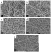
SEM analysis of neat PCL electrospun fibers obtained by setting different values of relative humidity (RH) : 25% (a), 30% (b), 40% (c), 50% (d) and 60% (e). Common scale bar 1 μm, higher magnification micrographs in the insets with common scale bar of 200 nm.
Figure 6
Figure 6
SEM/EDS analysis of the composite electrospun mats. SEM with maps and details for C (polymer component) and Si (from particles) (a) showing also the EDS spectrum on the particle cluster (b).
Figure 7
Figure 7
SEM micrograph of composite PCL_Ca_MCM-41 electrospun fibers after the tensile test, indicating lack of strong adhesion at the particle-PCL interface (arrows).
Figure 8
Figure 8
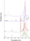
ATR-FTIR spectra of neat PCL electrospun fibers (a), PCL_Ca_MCM-41 composite electrospun fibers (b), the subtraction spectrum (neat PCL fibers subtracted from the composite fiber spectrum, (c) and the Ca_MCM-41 particle spectrum (d). Peaks of relevance are discussed in the text.
Figure 9
Figure 9
SEM micrograph of PCL_Ca_MCM-41 electrospun mats after 1 day (a) and 7 days (b) of immersion in SBF solution. Scale bar 1 μm and for the inset scale bar 200 nm.
Figure 10
Figure 10
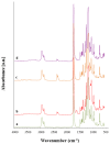
ATR-FTIR spectra of neat PCL fibers (a) and composite PCL_Ca_MCM-41 fibers (b) before immersion in SBF solution and PCL_Ca_MCM-41 fibers after immersion in SBF solution for 1 day (c) and 7 days (d).
Figure 11
Figure 11
Cell viability measured by using WST-8 assay on seeded electrospun samples of neat PCL microfibers, neat PCL nanofibers and composite PCL_Ca_MCM-41 fibers.
Figure 12
Figure 12
SEM micrographs of ST-2 cells seeded onto PCL_Ca_MCM-41 composite electrospun mats at different magnifications: 200× (a) and 10,000× (b).

References

    1. Kai D., Liow S.S., Loh X.J. Biodegradable polymers for electrospinning: Towards biomedical applications. Mater. Sci. Eng. C. Mater. Biol. Appl. 2014;45:659–670. doi: 10.1016/j.msec.2014.04.051. - DOI - PubMed
    1. Teo W.-E., Inai R., Ramakrishna S. Technological advances in electrospinning of nanofibers. Sci. Technol. Adv. Mater. 2011;12:013002. doi: 10.1088/1468-6996/12/1/013002. - DOI - PMC - PubMed
    1. Liu M., Duan X.-P., Li Y.-M., Yang D.-P., Long Y.-Z. Electrospun nanofibers for wound healing. Mater. Sci. Eng. C. 2017;76:1413–1423. doi: 10.1016/j.msec.2017.03.034. - DOI - PubMed
    1. Prabhakaran M.P., Venugopal J.R., Chyan T.T., Hai L.B., Chan C.K., Lim A.Y., Ramakrishna S. Electrospun biocomposite nanofibrous scaffolds for neural tissue engineering. Tissue Eng. Part A. 2008;14:1787–1797. doi: 10.1089/ten.tea.2007.0393. - DOI - PubMed
    1. Shin S.-H., Purevdorj O., Castano O., Planell J.A., Kim H.-W. A short review: Recent advances in electrospinning for bone tissue regeneration. J. Tissue Eng. 2012;3:2041731412443530. doi: 10.1177/2041731412443530. - DOI - PMC - PubMed

LinkOut - more resources