. 2017 Dec 20;9(12):736.
doi: 10.3390/polym9120736.

Immobilization of Platelet-Rich Plasma onto COOH Plasma-Coated PCL Nanofibers Boost Viability and Proliferation of Human Mesenchymal Stem Cells

Affiliations

Immobilization of Platelet-Rich Plasma onto COOH Plasma-Coated PCL Nanofibers Boost Viability and Proliferation of Human Mesenchymal Stem Cells

Anastasiya Solovieva et al. Polymers (Basel). .

Abstract

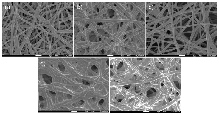
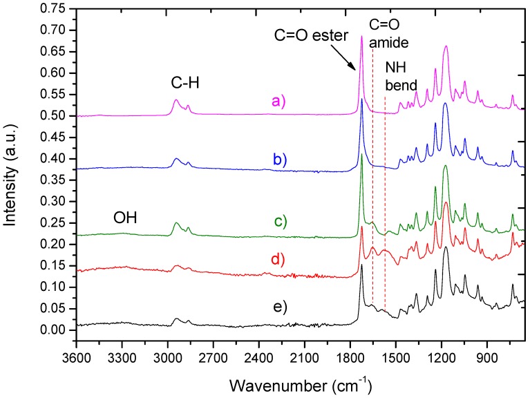
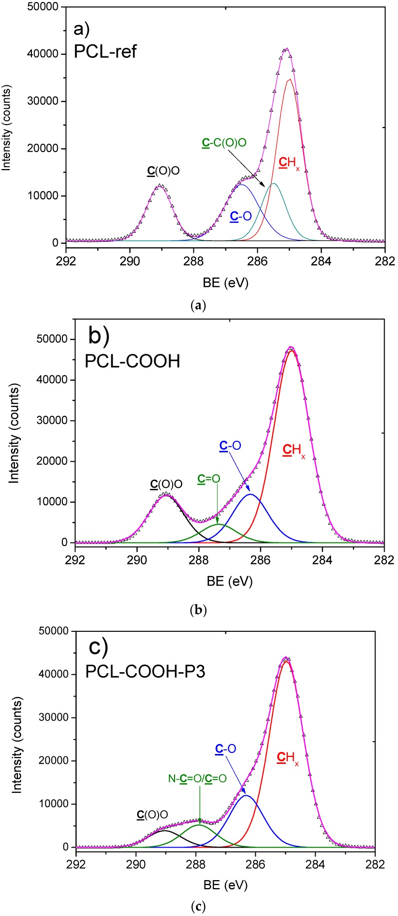
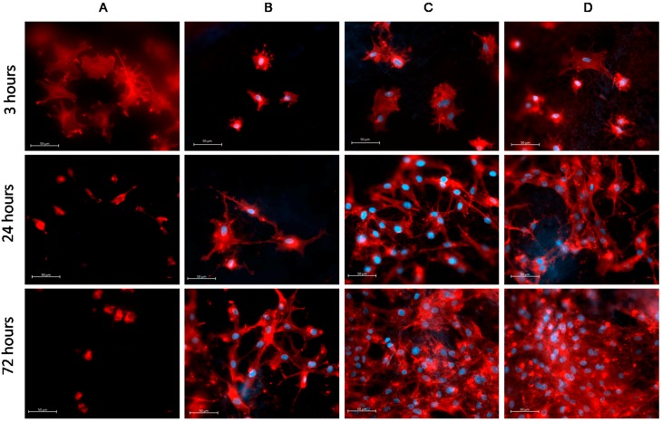
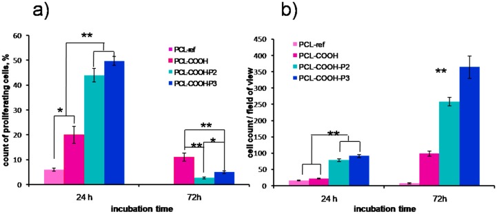
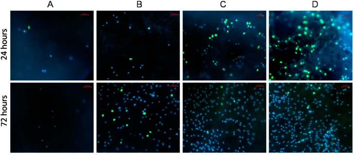
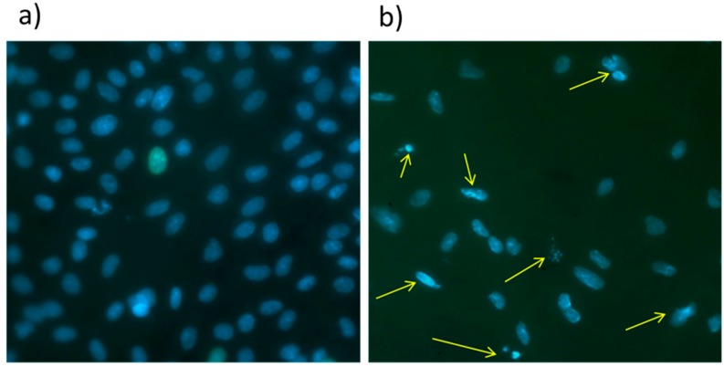
The scaffolds made of polycaprolactone (PCL) are actively employed in different areas of biology and medicine, especially in tissue engineering. However, the usage of unmodified PCL is significantly restricted by the hydrophobicity of its surface, due to the fact that its inert surface hinders the adhesion of cells and the cell interactions on PCL surface. In this work, the surface of PCL nanofibers is modified by Ar/CO₂/C₂H₄ plasma depositing active COOH groups in the amount of 0.57 at % that were later used for the immobilization of platelet-rich plasma (PRP). The modification of PCL nanofibers significantly enhances the viability and proliferation (by hundred times) of human mesenchymal stem cells, and decreases apoptotic cell death to a normal level. According to X-ray photoelectron spectroscopy (XPS), after immobilization of PRP, up to 10.7 at % of nitrogen was incorporated into the nanofibers surface confirming the grafting of proteins. Active proliferation and sustaining the cell viability on nanofibers with immobilized PRP led to an average number of cells of 258 ± 12.9 and 364 ± 34.5 for nanofibers with ionic and covalent bonding of PRP, respectively. Hence, our new method for the modification of PCL nanofibers with PRP opens new possibilities for its application in tissue engineering.

Keywords: COOH plasma; PRP immobilization; cell viability; nanofibers; platelet-rich plasma; polycaprolactone.


Conflict of interest statement

The authors declare no conflict of interest.

Figures

Figure 1
Figure 1
Scanning electron microscope (SEM) micrographs of polycaprolactone nanofibers (PCL-ref) (a); PCL-P1 (b); PCL-COOH (c); PCL-COOH-P2 (d) and PCL-COOH-P3 (e). The size of the bar corresponds to 1 µm.
Figure 2
Figure 2
Fourier transform infrared spectrophotometry using attenuated total reflectance mode (ATR-FTIR) spectra of as-prepared nanofibers PCL-ref (a); PCL-P1 (b); PCL-COOH (c); PCL-COOH-P2(d) and PCL-COOH-P3 (e).
Figure 3
Figure 3
X-ray photoelectron spectroscopy (XPS) C1s spectra of as-prepared nanofibers PCL-ref (a); PCL-COOH (b) and PCL-COOH-P3 (c).
Figure 4
Figure 4
Adhesion of mesenchymal stromal cells (MSCs) on the surface of PCL-ref (A); PCL-COOH (B); PCL-COOH-P2 (C) and PCL-COOH-P3 (D). The actin filaments of cytoskeleton are stained by Phalloidin (red) while the cell nucleus is stained by Hoechst 33342 (blue). All images shown at ×40 magnification and the size of the bar corresponds to 50 µm.
Figure 5
Figure 5
Influence of surface modification of PCL nanofibers on proliferation and viability of human MSCs. (a) the percentage of proliferating cells after 24 and 72 h of cultivation (calculated as the ratio of EdU-positive cells to the total number of Hoechst-positive cells); (b) average number of cells for single bright-field image after 24 and 72 h of cultivation. Data are expressed as mean ± standard deviation. ** p < 0.01, ** p < 0.05.
Figure 6
Figure 6
Influence of surface modification of PCL-ref (A); PCL-COOH (B); PCL-COOH-P2 (C) and PCL-COOH-P3 (D) nanofibers on proliferation and viability of human MSCs. The cell nucleus is stained by DNA binding fluorescent dye Hoechst 33342 (blue), the proliferating cells stained by EdUAlexa Fluor™ 488 (green). All images shown at ×20 magnification.
Figure 7
Figure 7
Representative picture of apoptosis analysis. The morphology of MSCs nucleus after 24 h. The PCL-COOH-P3 (a) exhibited homogenous form and coloration of the nucleus and no condensation of the chromatin; whereas cells grown on PCL-ref (b) exhibited the chromatin condensation and nuclear fragmentation (kariorhexis) (yellow arrows). Magnification ×40.

Similar articles

Cited by

References

    1. Yao Q., Cosme J.G.L., Xu T., Miszuk J.M., Picciani P.H.S., Fong H., Sun H. Three dimensional electrospun PCL/PLA blend nanofibrous scaffolds with significantly improved stem cells osteogenic differentiation and cranial bone formation. Biomaterials. 2017;115:115–127. doi: 10.1016/j.biomaterials.2016.11.018. - DOI - PMC - PubMed
    1. Woodruff M.A., Hutmacher D.W. The return of a forgotten polymer—Polycaprolactone in the 21st century. Prog. Polym. Sci. 2010;35:1217–1256. doi: 10.1016/j.progpolymsci.2010.04.002. - DOI
    1. Ghasemi-Mobarakeh L., Prabhakaran M.P., Morshed M., Nasr-Esfahani M.H., Ramakrishna S. Electrospun poly(ε-caprolactone)/gelatin nanofibrous scaffolds for nerve tissue engineering. Biomaterials. 2008;29:4532–4539. doi: 10.1016/j.biomaterials.2008.08.007. - DOI - PubMed
    1. Tokiwa Y., Calabia B.P., Ugwu C.U., Aiba S. Biodegradability of plastics. Int. J. Mol. Sci. 2009;10:3722–3742. doi: 10.3390/ijms10093722. - DOI - PMC - PubMed
    1. Feng Y., Liu W., Ren X., Lu W., Guo M., Behl M., Lendlein A., Zhang W. Evaluation of electrospun PCL-PIBMD meshes modified with plasmid complexes in vitro and in vivo. Polymers. 2016;8:1–14. doi: 10.3390/polym8030058. - DOI - PMC - PubMed