Review
. 2018 Apr 6;10(4):410.
doi: 10.3390/polym10040410.

Development of Mucoadhesive Chitosan Derivatives for Use as Submucosal Injections

Affiliations
Review

Development of Mucoadhesive Chitosan Derivatives for Use as Submucosal Injections

Hidemi Hattori et al. Polymers (Basel). .

Abstract

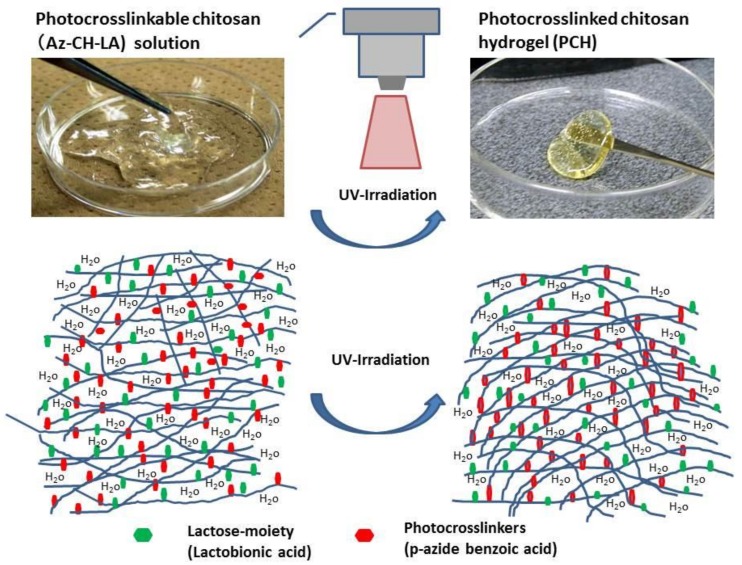
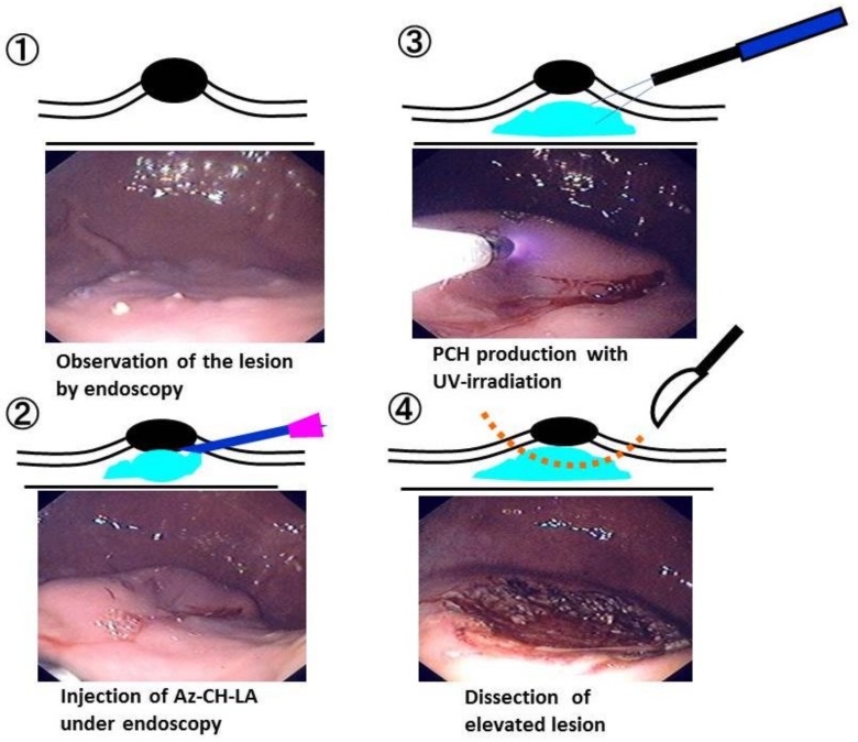
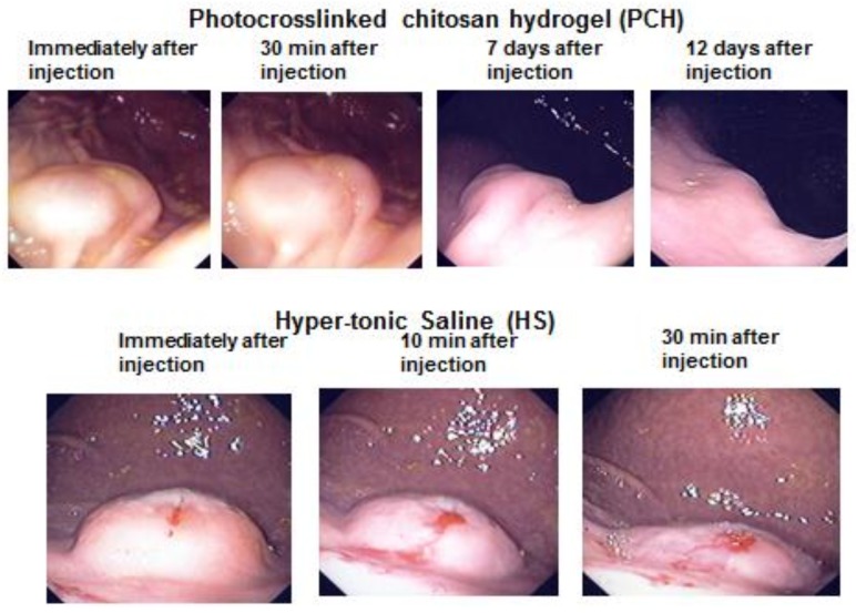
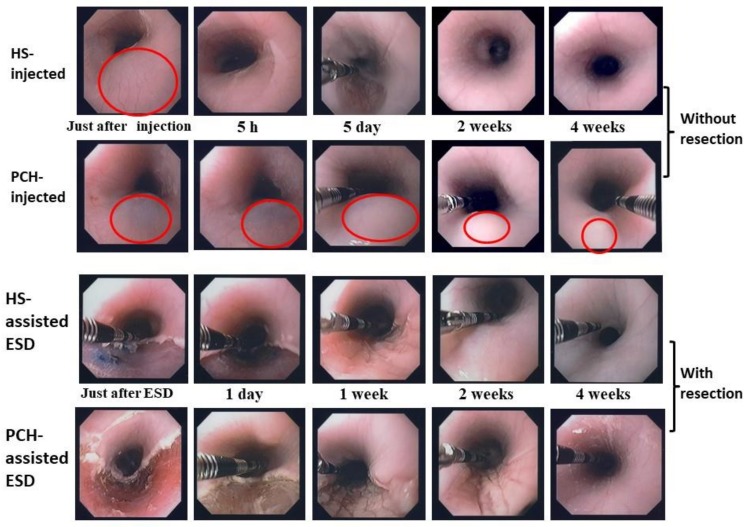
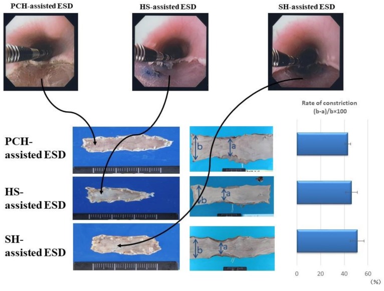
Endoscopic mucosal resection (EMR) and endoscopic submucosal dissection (ESD) have been used for surgical treatment of early gastric cancer. These endoscopic techniques require proper submucosal injections beneath the tumor to provide a sufficiently high submucosal fluid cushion (SFC) to facilitate clean dissection and resection of the tumor. Until now, the submucosal injection materials developed for endoscopic techniques such as EMR and ESD of tumors have been composed of macromolecules, proteins, or polysaccharides. We have been investigating the use of chitosan, a product that is obtained by the alkaline deacetylation of chitin, the second-most abundant natural polysaccharide. Specifically, we have been studying a photocrosslinked chitosan hydrogel (PCH) and solubilized chitosan derivatives for use as novel submucosal injections for endoscopic techniques. Notably, chitosan derivatives with lactose moieties linked to the amino groups of its glucosamine units can specifically interact with acidic mucopolysaccharides and mucins in submucosa without the need for the incorporation of harmful photoreactive groups nor potentially mutagenic ultraviolet irradiation.

Keywords: chitosan derivatives; endoscopic mucosal resection; endoscopic submucosal dissection; mucoadhesive; mucopolysaccharide; submucosal injection.


Conflict of interest statement

The authors declare no conflict of interest.

Figures

Figure 1
Figure 1
Photocrosslinkable chitosan (Az-CH-LA) solution and photocrosslinked chitosan hydrogel (PCH). The viscous Az-CH-LA solution (upper, left photograph) was converted to insoluble soft rubber-like PCH (upper, right photograph) through crosslinking reaction of the azide and amino groups of Az-CH-LA with UV-irradiation (lower illustration).
Figure 2
Figure 2
Representative images of Az-CH-LA injection, ultraviolet (UV) irradiation, and the appearance when PCH was used as SFC in PCH-assisted ESD. ① Observation of the lesion by endoscopy, ② Injection of Az-CH-LA under endoscopy, ③ PCH production with UV-irradiation, ④ Dissection of elevated lesion. These experiments demonstrated the feasibility and safety of PCH-assisted ESD in a porcine stomach model. Reprinted from [5], Copyright 2009, with permission from Thieme.
Figure 3
Figure 3
Endoscopic appearance at injection sites of PCH or HS. The change of each injection site was observed endoscopically, without dissection, at the indicated time points. Reprinted from [5], Copyright 2009, with permission from Thieme.
Figure 4
Figure 4
Changes in endoscopic appearance of hypertonic saline (HS)- or PCH-injected sites of pig esophagus (in live animals) without (upper 2 rows) and with (lower 2 rows) mucosal resection. Endoscopic changes of mucosal elevation shapes are shown at the following times after the injection of 2 mL HS or PCH: just after injection, and 5 h, 5 days, 2 weeks, and 4 weeks later. Endoscopic appearances are shown at the following times after HS- and PCH-assisted ESD: just after ESD, and 1 day, 1 week, 2 weeks, and 4 weeks later. Note the duration of elevation in the PCH-injected case (2nd row vs. 1st row; red circles represent centers of mucosal elevation). Reprinted from [7], Copyright 2012, with permission from Elsevier.
Figure 5
Figure 5
Endoscopic appearance following endoscopic submucosal dissection (ESD). (Upper panel): Endoscopic appearance immediately after ESD assisted by photocrosslinkable chitosan hydrogel (PCH), hypertonic saline solution (HS), and sodium hyaluronate (SH) in pig esophagus in living animals (left to right, respectively). (Lower panel): Left column: Resected tissues recovered from pig esophagi in each group, showing constriction of esophagus following ESD assisted by PCH, HS, and SH. Middle column: Resected tissues recovered from inner esophagus at 4 weeks after ESD. Right column: Calculated rate of constriction (%; mean ± SD, n = 6) at 4 weeks after ESD. Photographs are representative images from the indicated groups. Reprinted from [7], Copyright 2012, with permission from Elsevier.
Figure 6
Figure 6
Lifting effect of CH-LA-S. A solution of 3% CH-LA-S (black columns), 2% CH-LA-S (gray columns), or physiological saline (white columns) was injected into the submucosa of ex vivo porcine stomach, and the heights of injection volumes were measured at the indicated time point over the subsequent hour. Data are plotted as mean ± SD (n = 5). *p < 0.05, **p < 0.01, ***p < 0.005, and ****p < 0.001 by Tukey post-hoc test. Reprinted from [27], Copyright 2017, with permission from Elsevier.
Figure 7
Figure 7
(A): Scheme of when CH-LA-S is injected into submucosa. CH-LA-S is adhered to parts in contact with tissues, but the inside of the injected CH-LA-S remains fluid. (B): Scheme of different formation patterns due to difference in amount of CH-LA-S and sulfated mucopolysaccharides. Reprinted from [27], Copyright 2017, with permission from Elsevier.

Similar articles

Cited by

References

    1. Uedo N., Takeuchi Y., Ishihara R. Endoscopic management of early gastric cancer: Endoscopic mucosal resection or endoscopic submucosal dissection: Data from a Japanese high-volume center and literature review. Ann. Gastroenterol. 2012;25:281–290. - PMC - PubMed
    1. Shim C.S. Endoscopic mucosal resection: An overview of the value of different techniques. Endoscopy. 2001;33:271–275. doi: 10.1055/s-2001-12816. - DOI - PubMed
    1. Ning B., Abdelfatah M.M., Othman M.O. Endoscopic submucosal dissection and endoscopic mucosal resection for early stage esophageal cancer. Ann. Cardiothorac. Surg. 2017;6:88–98. doi: 10.21037/acs.2017.03.15. - DOI - PMC - PubMed
    1. Fujishiro M., Yahagi N., Kashimura K., Matsuura T., Nakamura M., Kakushima N., Kodashima S., Ono S., Kobayashi K., Hashimoto T., et al. Tissue damage of different submucosal injection solutions for EMR. Gastrointest. Endosc. 2005;62:933–942. doi: 10.1016/j.gie.2005.07.052. - DOI - PubMed
    1. Ishizuka T., Ishihara M., Aiko S., Nogami Y., Nakamura S., Kanatani Y., Kishimoto S., Hattori H., Horio T., Tanaka Y., et al. Experimental evaluation of photocrosslinkable chitosan hydrogel as injection solution for endoscopic resection. Endoscopy. 2009;41:25–28. doi: 10.1055/s-0028-1103483. - DOI - PubMed

LinkOut - more resources