Review
. 2018 Apr 11;10(4):428.
doi: 10.3390/polym10040428.

Aliphatic Polyester Nanofibers Functionalized with Cyclodextrins and Cyclodextrin-Guest Inclusion Complexes

Affiliations
Review

Aliphatic Polyester Nanofibers Functionalized with Cyclodextrins and Cyclodextrin-Guest Inclusion Complexes

Ganesh Narayanan et al. Polymers (Basel). .

Abstract

The fabrication of nanofibers by electrospinning has gained popularity in the past two decades; however, only in this decade, have polymeric nanofibers been functionalized using cyclodextrins (CDs) or their inclusion complexes (ICs). By combining electrospinning of polymers with free CDs, nanofibers can be fabricated that are capable of capturing small molecules, such as wound odors or environmental toxins in water and air. Likewise, combining polymers with cyclodextrin-inclusion complexes (CD-ICs), has shown promise in enhancing or controlling the delivery of small molecule guests, by minor tweaking in the technique utilized in fabricating these nanofibers, for example, by forming core⁻shell or multilayered structures and conventional electrospinning, for controlled and rapid delivery, respectively. In addition to small molecule delivery, the thermomechanical properties of the polymers can be significantly improved, as our group has shown recently, by adding non-stoichiometric inclusion complexes to the polymeric nanofibers. We recently reported and thoroughly characterized the fabrication of polypseudorotaxane (PpR) nanofibers without a polymeric carrier. These PpR nanofibers show unusual rheological and thermomechanical properties, even when the coverage of those polymer chains is relatively sparse (~3%). A key advantage of these PpR nanofibers is the presence of relatively stable hydroxyl groups on the outer surface of the nanofibers, which can subsequently be taken advantage of for bioconjugation, making them suitable for biomedical applications. Although the number of studies in this area is limited, initial results suggest significant potential for bone tissue engineering, and with additional bioconjugation in other areas of tissue engineering. In addition, the behaviors and uses of aliphatic polyester nanofibers functionalized with CDs and CD-ICs are briefly described and summarized. Based on these observations, we attempt to draw conclusions for each of these combinations, and the relationships that exist between their presence and the functional behaviors of their nanofibers.

Keywords: controlled drug delivery; cyclodextrin-inclusion complexes; cyclodextrins; mechanical properties; poly(lactic acid); poly(ε-caprolactone); pseudorotaxanes; rapid dissolution.


Conflict of interest statement

The authors declare no conflict of interest.

Figures

Figure 1
Figure 1
Schematic representations of the native cyclodextrins (CDs) (α-, β-, and γ-CDs). All three native CDs have a similar depth or height (7.9 Å); however, a key difference lies in their inner diameters, with α-, β- and γ-CDs having inner diameters of 5.7, 7.8, and 9.5 Å, respectively. Images adapted and reproduced from [11]. Copyright 2017 Elsevier Ltd.
Figure 2
Figure 2
Scanning Electron Micrographs (SEM) of PCL and PCL/γ-CD functionalized nanofibers. By simply adjusting the PCL concentration from 14% to 12%, an increase in γ-CD loading (from 15% to upwards of 30%) was made possible without significant increases in the beaded structures in the nanofibers (AC). Images adapted and reproduced with permission from [42]. Copyright American Chemical Society 2014.
Figure 3
Figure 3
Water Contact Angle (WCA) values of PCL, PCL/α-CD, and PCL/γ-CD functionalized nanofibers. The neat PCL showed hydrophobic characteristics (WCA of 140°), while the WCA of functionalized nanofibers showed a marked decrease in hydrophobic characteristics, even with the addition of as little as 5% CD (WCA of PCL/5% α-CD and PCL/5% γ-CD are ~120°). With further addition, WCA plateaued at ~100°, depending on the CD used. Images adapted and reproduced with permission from [43]. Copyright American Chemical Society 2014.
Figure 4
Figure 4
Wide-angle diffraction (WAXD) patterns of the electrospun PCL, α-CD functionalized electrospun PCL nanofibers, and PCL-α-CD film cast from solution. Neat PCL showed two peaks at 22° and 24° 2θ corresponding to (110) and (200) reflections. While the lack of any additional peaks in 10% α-CD containing PCL nanofibers, the presence of an additional peak at 20° 2θ and shifts in the PCL peaks, indicate the presence of some inclusion complex between PCL and α-CD. Images adapted and reproduced from [42]. Copyright American Chemical Society 2014.
Figure 5
Figure 5
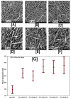
SEM images and average fiber diameters of β-CD functionalized PCL nanofibers at various β-CD loadings (0% to 50%). SEM images indicate the absence of beaded structures in PCL and PCL/β-CD nanofibers. While a marginal increase in fiber size was observed with the addition of β-CD (520 ± 200 nm for 10% β-CD vs. 400 ± 160 nm for neat PCL), further increases in β-CD concentration did not lead to further increases in fiber diameter. Images adapted and reproduced with permission from [43]. Copyright John Wiley and Sons 2015.
Figure 6
Figure 6
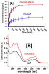
The release and solubility profiles of naproxen (NAP) from PCL and PCL/β-CD-IC nanofibers. The release profile study (A) by HPLC revealed significant release of NAP from PCL/NAP-β-CD-IC, compared to the PCL/NAP nanofibers, owing to improved solubility of encapsulated NAP, compared to pristine NAP. These results were further corroborated by solubility analyses (UV-vis) (B), which showed significantly enhanced solubility, as observed by increases in the intensity. Images adapted and reproduced from [63]. Copyright Elsevier Ltd., 2014.
Figure 7
Figure 7
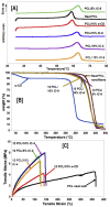
Structure–property relationships (thermal and mechanical), due to the presence of intact non-stoichiometric inclusion complexes (n-s PCL-α-CD-IC) in PCL nanofibers. The presence of intact (n-s PCL-α-CD-IC) caused increases (~5 °C) in PCL melting temperature (A), irrespective of the stoichiometric ratio of the IC or the wt % loading of those ICs in the nanofibers. Likewise, the degradation temperature of the α-CD phase significantly increased in those composites, indicating the presence of α-CD in complexed state (B). Finally, intact ICs caused changes in the mechanical behavior (increased modulus value and decreased elongation) of the composites. Images adapted with permission from [95]. Copyright Elsevier Ltd., 2015.
Figure 8
Figure 8
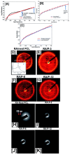
Establishing structure property relationships between pseudorotaxane (PpR) solution rheology and the resultant molecular arrangement of PpR in the nanofibers. Solution rheology of neat PCL solutions at higher concentrations (12%) showed low modulus values (elastic and loss) with multiple cross-over points, typical of viscoelastic material. On the other hand, PpR solutions showed frequent cross-over points, indicating a Rouse–Zimm effect (AC). However, both 2D-wide angle X-ray diffraction (DG) and selected area electron diffraction analyses (HK) showed the presence of crystalline domains in both randomly aligned, as well as aligned PCL nanofibers, while no such peaks were evidently observed in PpR nanofibers. Images adapted and reproduced with permission from [97]. Copyright The Royal Society of America 2016.
Figure 9
Figure 9
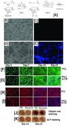
Schematic illustrating the immobilization of fluorescent molecules and the effect of hydroxyl groups of cyclodextrins on the osteogenic differentiation of human adipose-derived stem cells. The Neat PCL does not have surface epitopes facilitating immobilization of bioactive molecules. Conversely, the pseudorotaxanes contain abundant hydroxyl groups, facilitating the immobilization of bioactive molecules, and a representative fluorescent molecule (fluorescamine) (A), whose presence was monitored by fluorescent spectroscopy (BE). Although immobilization of active biomolecule was possible only in scaffolds containing CDs, live-dead cell assay and F-actin assay showed no statistical significance in the cell viability at any of the time point studied (FI) In addition to facilitating the immobilization of bioactive molecules, PCL/α-CD nanofibers without immobilization of biomolecules also facilitated the osteogenic differentiation of human adipose derived stem cells (h-ADSCs), although the osteogenic marker levels showing similarities in the expression levels between the study groups were the same (J,K). Images adapted and reproduced with permission from [103]. Copyright Springer 2012.
Figure 10
Figure 10
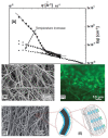
Temperature-dependent stability of pseudorotaxanes (PpR) using DMSO and an electron micrograph of electrospun core–shell nanofibers with PpR in the shell region, which was subsequently conjugated with FITC. The SANS analyses of the PpR (shell region) in DMSO showed decreases in scattering intensity, indicating dethreading of PpRs into α-CD and PCL phases. Whereas at 35 °C, higher scattering intensity indicated the presence of a PpR structure, and in addition, a parallelepiped model was developed which showed CD assemblies approximating 11 α-CDs in length and 20 α-CDs in width (A). Additionally, conjugation of fluorescamine was only possible onto hydroxyl groups present in PpR (BE), whereas in neat PCL nanofibers, conjugation was not possible. Images adapted and reproduced with permission from [104]. Copyright John Wiley and Sons 2015.
Figure 11
Figure 11
Proliferation and cell densities of Michigan Cancer Foundation-7 (MCF-7) cells on poly(lactide-co-caprolactone) (PLCL or PLACL) scaffolds and PLCL scaffolds containing aloe vera (AV), magnesium oxide (MgO), and free curcumin (CUR), or encapsulated in β-CD. The addition of AV alone did not cause MCF-7 cell death at all studied time points; however, the addition of CUR, in either free or encapsulated form, resulted in significant cell death of the proliferating MCF-7 cells (A). These results were further corroborated by laser confocal scanning microscopy measurements, which showed low cell densities in scaffolds containing CUR and MgO/CUR (11 B: (E,F)). Images adapted and reproduced with permission from [109]. Copyright Elsevier Ltd., 2017.
Figure 12
Figure 12
The effects of poly(lactic acid) nanofibers containing β-CD/cinnamaldehyde (CA) inclusion complex on the cumulative release of CA and cell viability of human skin fibroblasts. The cumulative release of CA depended on the β-CD/CA concentration, i.e., the higher the concentration, the higher the release rate of CA, possibly owing to improved dissolution of CA from β-CD complex (A). In comparison, low cell densities of S. aureus and E. coli were observed with increasing β-CD/CA complex concentrations. Similar trends were also seen with the proliferation of human skin fibroblasts cells, when exposed to CA containing PLA nanofibers or free CA (B). Images adapted and reproduced with permission from [111] Copyright Liu et al., 2017.
Figure 12
Figure 12
The effects of poly(lactic acid) nanofibers containing β-CD/cinnamaldehyde (CA) inclusion complex on the cumulative release of CA and cell viability of human skin fibroblasts. The cumulative release of CA depended on the β-CD/CA concentration, i.e., the higher the concentration, the higher the release rate of CA, possibly owing to improved dissolution of CA from β-CD complex (A). In comparison, low cell densities of S. aureus and E. coli were observed with increasing β-CD/CA complex concentrations. Similar trends were also seen with the proliferation of human skin fibroblasts cells, when exposed to CA containing PLA nanofibers or free CA (B). Images adapted and reproduced with permission from [111] Copyright Liu et al., 2017.
Figure 13
Figure 13
Total cumulative release and antioxidant characteristics of gallic acid (GA) released from PLA/GA-HP-β-CD nanofibers. The total cumulative release of GA from PLA/GA-HP-β-CD nanofibers was always higher in water (A) and 10% ethanol solutions (B), than those released from PLA/GA nanofibers, owing to the enhanced dissolution characteristics of GA/HP-β-CD compared to free GA. In addition, the DPPH assay showed that the presence of GA in both PLA/GA, as well as PLA/GA-HP-β-CD nanofibers, retained its antioxidant characteristics (C). Images adapted and reproduced with permission from [114]. Copyright Elsevier Ltd., 2016.

References

    1. Kny E., Uyar T., Kny E. In: Electrospun Materials for Tissue Engineering and Biomedical Applications. Kny E., Uyar T., editors. Woodhead Publishing; Sawston, UK: 2017. pp. i–iii.
    1. Anu Bhushani J., Anandharamakrishnan C. Electrospinning and electrospraying techniques: Potential food based applications. Trends Food Sci. Technol. 2014;38:21–33. doi: 10.1016/j.tifs.2014.03.004. - DOI
    1. Wang W., Huang H., Li Z., Zhang H., Wang Y., Zheng W., Wang C. Zinc Oxide Nanofiber Gas Sensors via Electrospinning. J. Am. Ceram. Soc. 2008;91:3817–3819. doi: 10.1111/j.1551-2916.2008.02765.x. - DOI
    1. Narayanan G., Vernekar V.N., Kuyinu E.L., Laurencin C.T. Poly (lactic acid)-based biomaterials for orthopaedic regenerative engineering. Adv. Drug Deliv. Rev. 2016;107:247–276. doi: 10.1016/j.addr.2016.04.015. - DOI - PMC - PubMed
    1. Ramakrishna S., Fujihara K., Teo W.-E., Yong T., Ma Z., Ramaseshan R. Electrospun nanofibers: Solving global issues. Mater. Today. 2006;9:40–50. doi: 10.1016/S1369-7021(06)71389-X. - DOI

LinkOut - more resources